Properties and uses of chaga
text_fieldstext_fieldsarrow_upward
Pharmacological properties of chaga
Chaga
- increases the body's defense reactions in the experiment,
- activates the metabolism in the brain tissue, which is manifested by an increase in the bioelectrical activity of the cerebral cortex.
- acts anti-inflammatory with
- internal and
- topical application.
In experimental studies of chaga
inhibits the growth of some tumors.
The use of chaga
enhances the cytostatic effects of cyclophosphamide.
Birch mushroom decoction
has a hypoglycemic effect: the maximum decrease in serum glucose levels is observed 1.5-3 hours after ingestion of the decoction. The sugar level drops by 15.8-29.9%.
It is noted that a decoction from the inner part of the mushroom gives a hypoglycemic effect, a decoction from the outer part does not possess this property.
The use of chaga
Chaga is used as
- fortifying and
- anti-inflammatory agent for diseases of the gastrointestinal tract and
- as a symptomatic remedy for tumors of various localization.
Chaga infusion is prescribed to patients
peptic ulcer of the stomach and duodenum.
Chaga
- quickly relieves pain and dyspeptic symptoms,
- normalizes bowel function,
- increases overall tone.
The positive effect of chaga on patients with gastrointestinal diseases is confirmed by X-ray data.
Infusion and tincture of chaga, "Befungin" is used for
- psoriasis,
- eczema
- and other skin diseases,
the treatment is especially effective in cases of a combination of a skin disease with various inflammatory diseases of the gastrointestinal tract, liver, and biliary system.
When treating chaga, the patient is recommended to eat mainly dairy and vegetable food, to limit the intake of meat and fats, exclude canned food, smoked meats, hot spices. It is also impossible to inject glucose intravenously and use penicillin.
Rhodotus palmatus (Rhodotus palmatus)
Other names
- Dendrosarcus subpalmatus;
- Pleurotus subpalmatus;
- Gyrophila palmata;
- Rhodotus subpalmatus.
The palm-shaped Rhodotus is the only member of the genus Rhodotus, belonging to the Physalacriaceae family, and has a rather specific appearance. The pink or pinkish-orange cap of this fungus in mature fruiting bodies is densely speckled with a venous mesh. Because of this appearance, the described mushroom is often called a shriveled peach. The fruity aroma of the mushroom pulp contributed to the emergence of this name to some extent. The palatability of the palm-shaped rhodotus is not very good, the pulp is very bitter, elastic.
External description
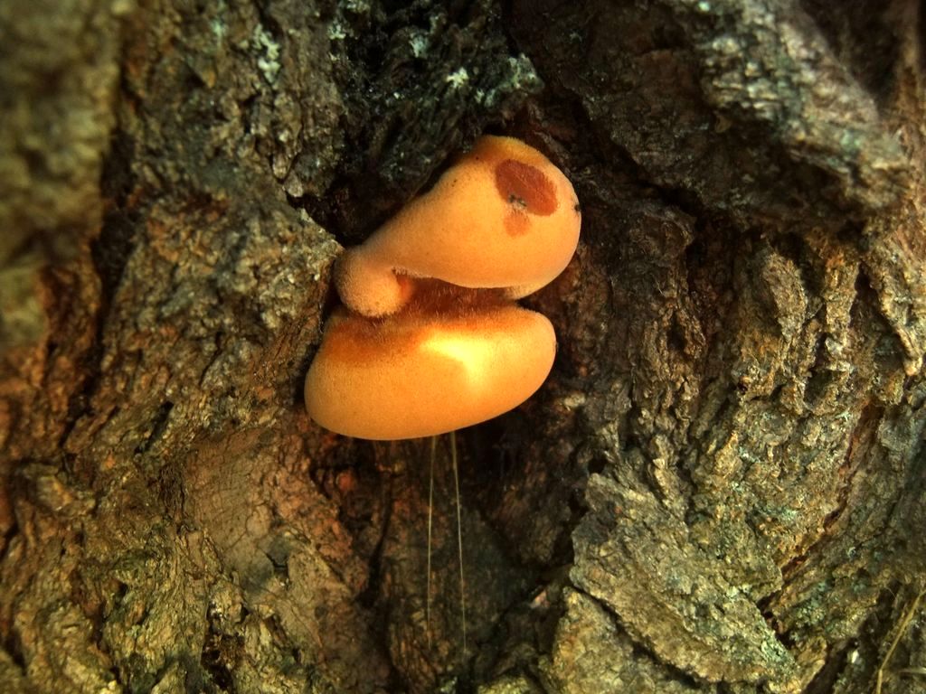
The fruiting body of the palm-shaped rhodotus is hat-pectus. The mushroom cap has a diameter of 3-15 cm, a convex shape and a curved edge, very elastic, initially with a smooth surface, and in old mushrooms it is covered with a wrinkled venous mesh. Only sometimes the surface of the cap of this mushroom remains unchanged. The mesh appearing on the cap of the mushroom is slightly lighter in color than the rest of the surface, while the color of the cap between the wrinkled scars may change. The color of the surface will depend on how intense the lighting was during the development of the fruiting body of the fungus. It can be orange, salmon or pink. In young mushrooms, the fruiting body can secrete droplets of reddish liquid.
The leg of the mushroom is located in the center, more often - eccentric, has a length of 1-7 cm, and in diameter is 0.3-1.5 cm, sometimes hollow, the flesh of the leg is very hard, has a small edge on its surface, pinkish, but without a volva and a peri-cap ring ... The length of the leg will depend on how high-quality the illumination of the fruiting body was during the period of its development.
The mushroom flesh of the palm-shaped rhodotus is elastic, has a jelly-like layer located under the thin skin of the cap, a bitter taste and a barely pronounced fruity aroma, reminiscent of the smell of citrus fruits or apricots. When interacting with iron salts, the color of the pulp immediately changes, becoming dark green.
The hymenophore of the described fungus is lamellar.Elements of the hymenophore - plates, located freely, can be creeping along the stem of the fungus or notched-attached. Often they have an abdomen, greater thickness and frequency of location. Moreover, large plates of the hymenophore are often interspersed with small and thin ones. According to the color of the plate of the described fungus, they are pale-salmon-pink, some of them do not reach the edge of the cap and the base of the leg. Fungal spores are 5.5-7 * 5-7 (8) microns in size. Their surface is covered with warts, and the spores themselves are often spherical.
Season and habitat of the mushroom
Rhodotus palmatus belongs to the saprotroph category. He prefers to live mainly on stumps and trunks of deciduous trees. Occurs singly or in small groups, mainly on elm fallen trees. There is information about the growth of the described species of mushrooms on the wood of maple, American linden, horse chestnut. Griyu rhodotus palmate is widespread in many European countries, in Asia, North America, New Zealand, Africa. In mixed coniferous and deciduous forests, such mushrooms can be seen very rarely. Active fruiting of the palm-shaped rhodotus occurs from spring to late autumn.
Edibility
The palm-shaped rhodotus (Rhodotus palmatus) is inedible. In general, its nutritional properties have been little studied, but too hard pulp does not allow eating this mushroom. Actually, these properties of the pulp make the described type of mushrooms inedible.
Similar types and differences from them
The palm-shaped rhodotus has a rather specific appearance. The cap in young mushrooms of this species is pinkish, and in mature ones it is orange-pink, and on its surface, a net of thin and closely intertwined veins characteristic of this species is almost always visible. Such signs do not allow the described mushroom to be confused with any other, moreover, the pulp of the fruiting body has a clearly distinguishable fruity aroma.
Other information about the mushroom
Despite the fact that the Rhodotus palmate belongs to the number of inedible mushrooms, it has some medicinal properties. They were discovered in 2000 by a group of Spanish microbiologists. Research has confirmed that this type of fungus has good antimicrobial activity against human pathogens.
Rhodotus palmatus is included in the Red Book of several countries (Austria, Estonia, Romania, Poland, Norway, Germany, Sweden, Slovakia).
General information about bristly tinder fungus
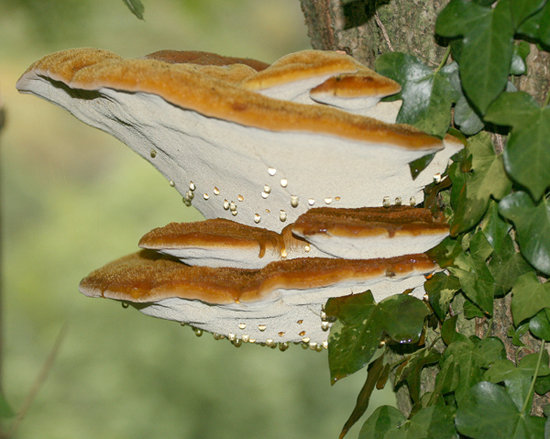
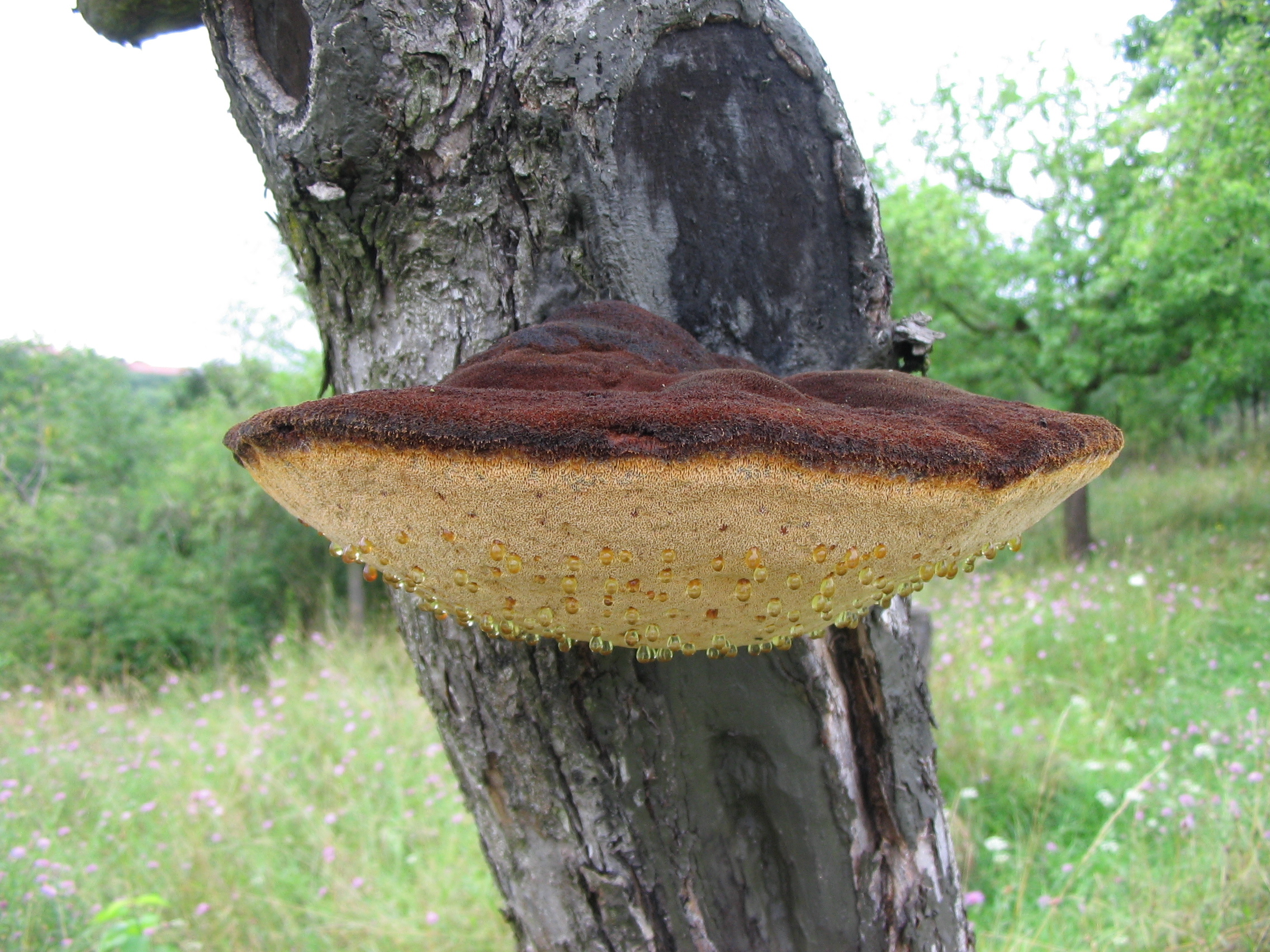
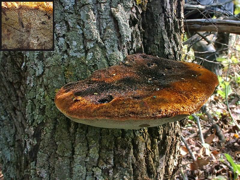
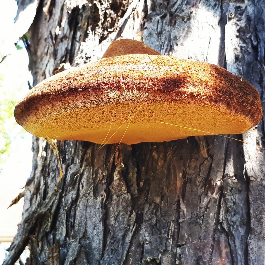
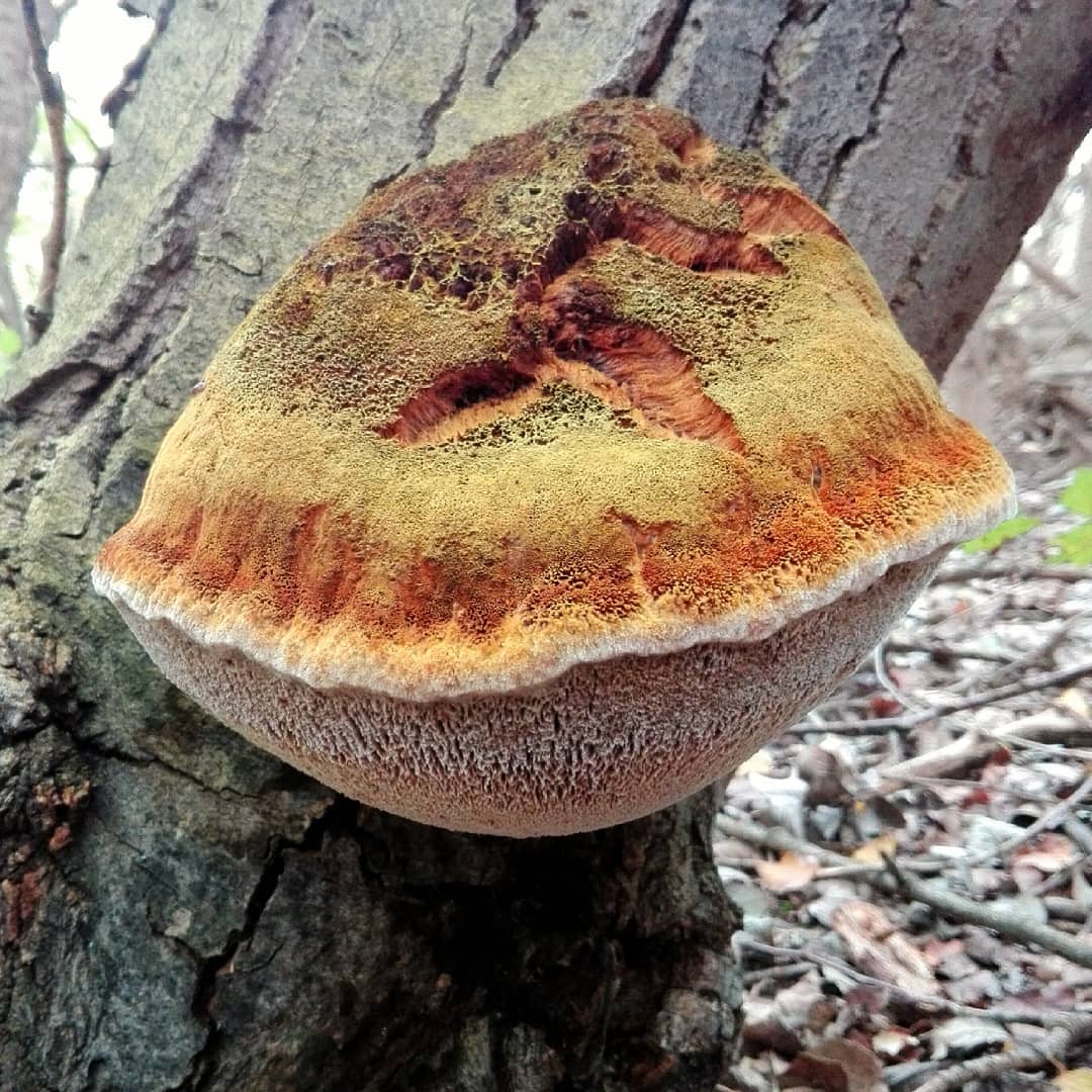
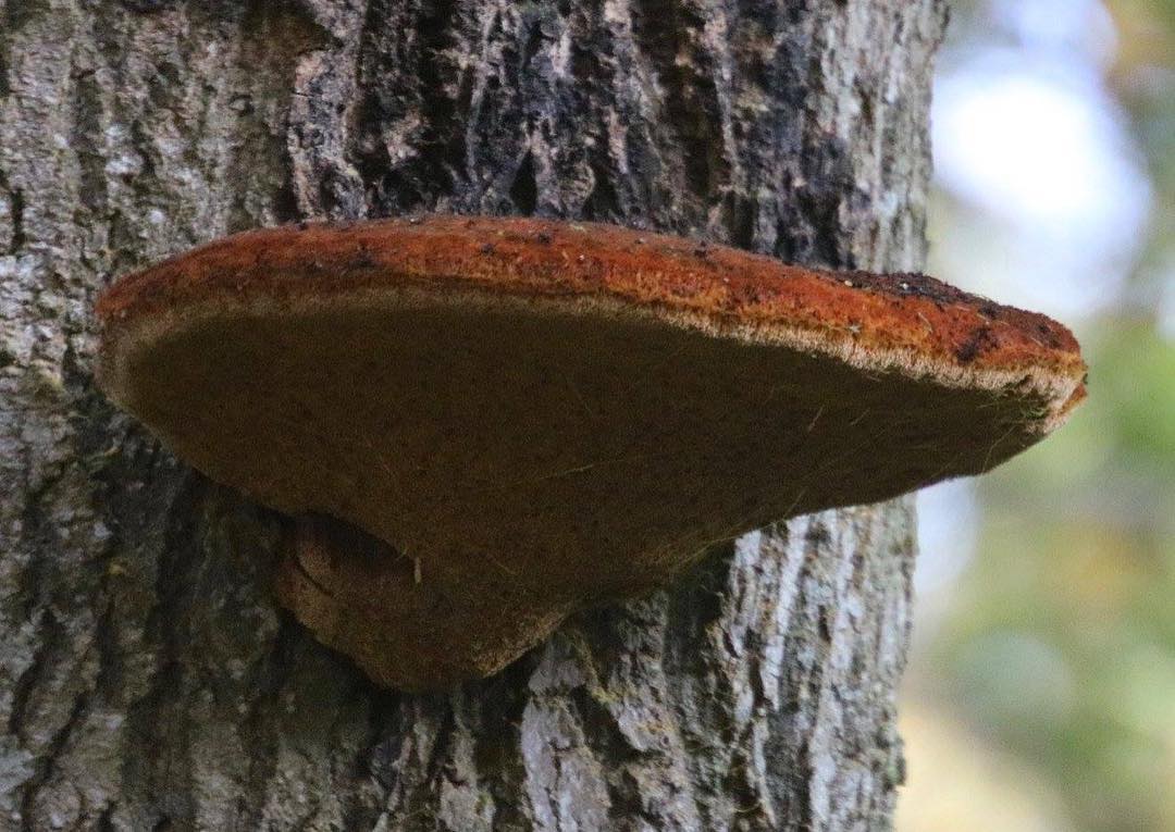
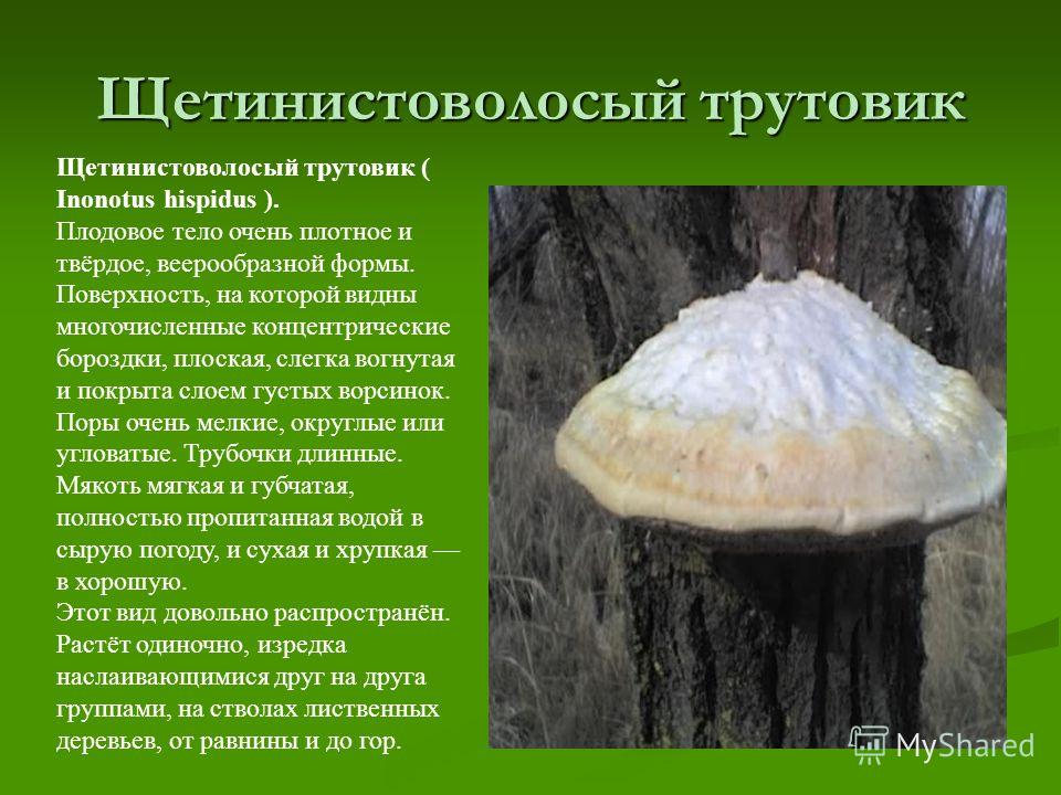
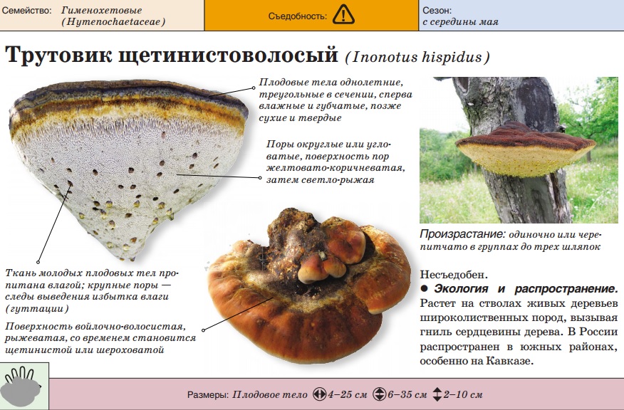
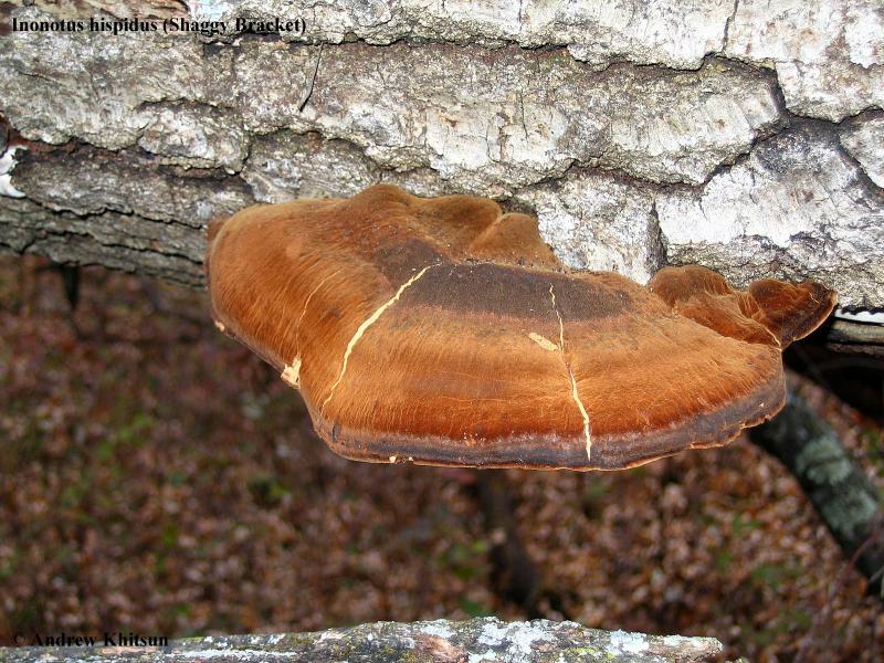
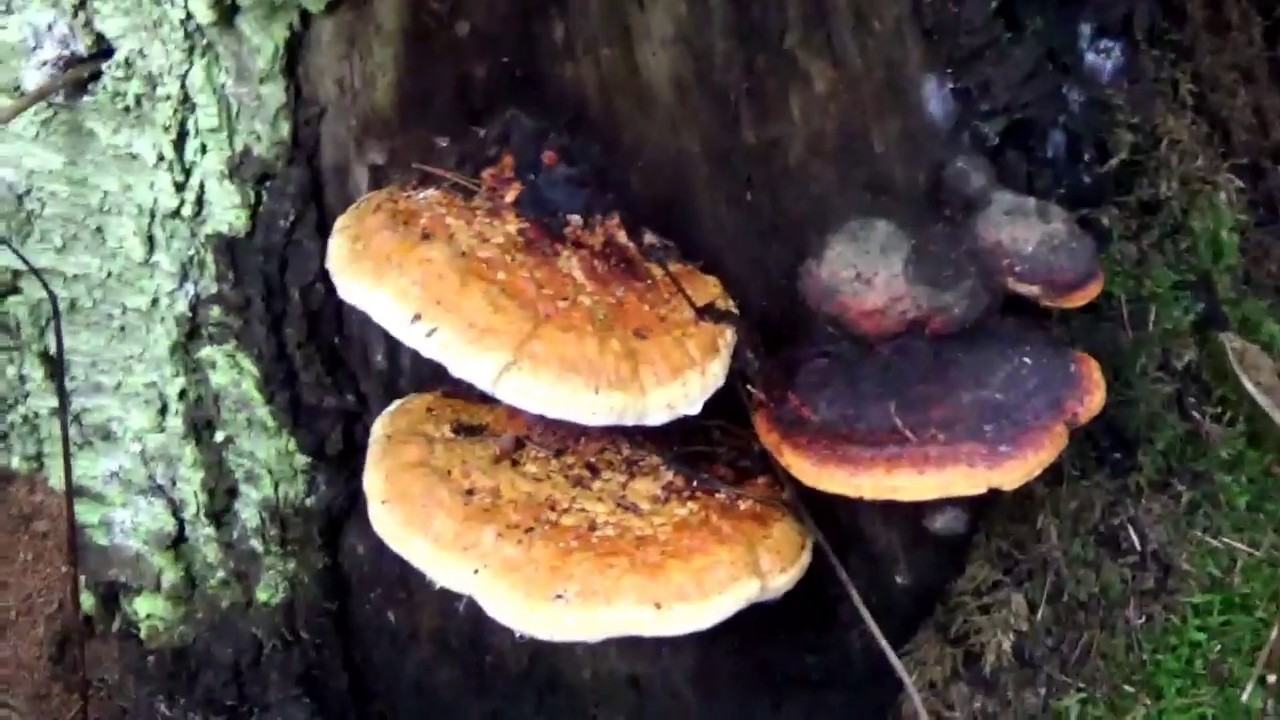
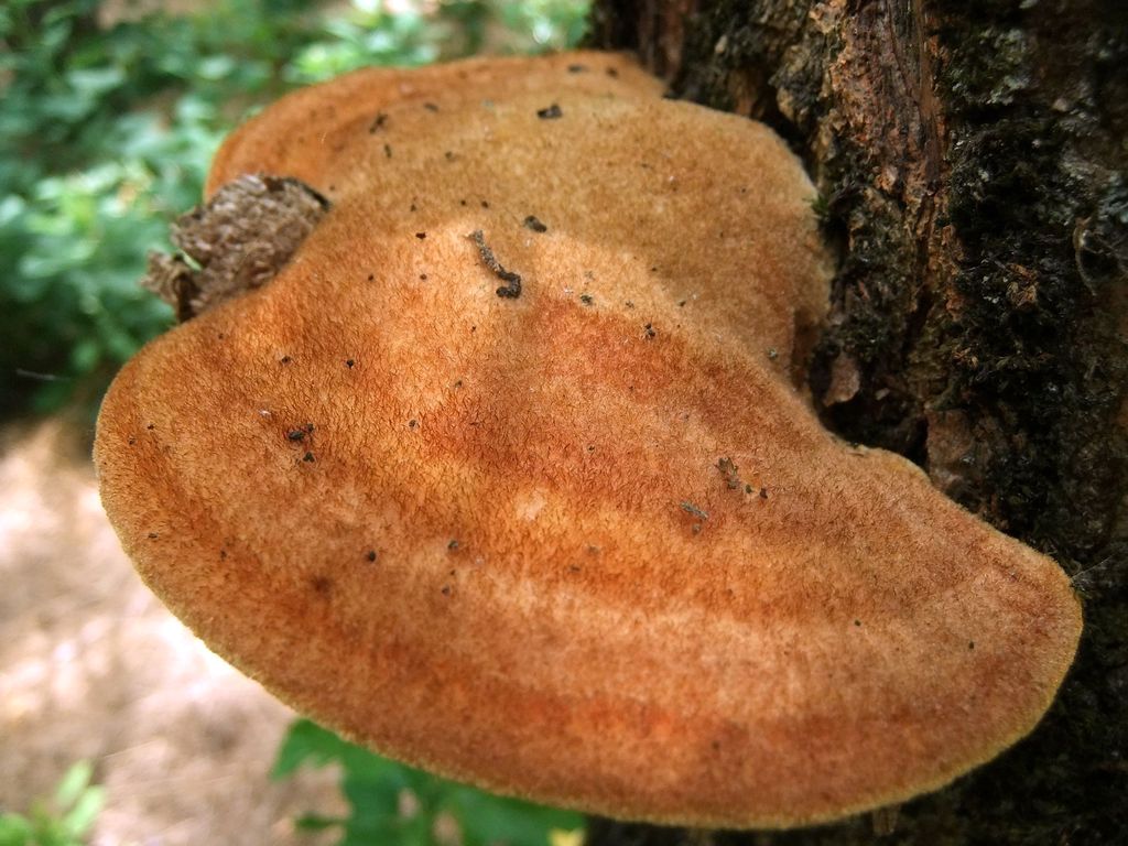
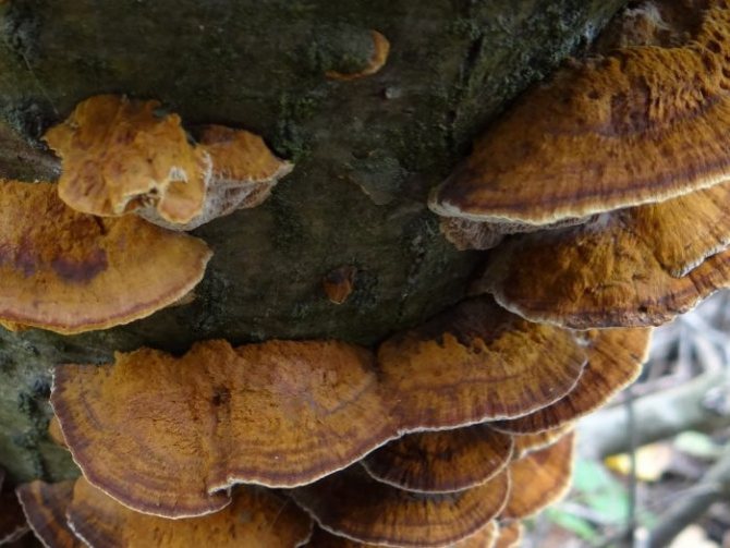
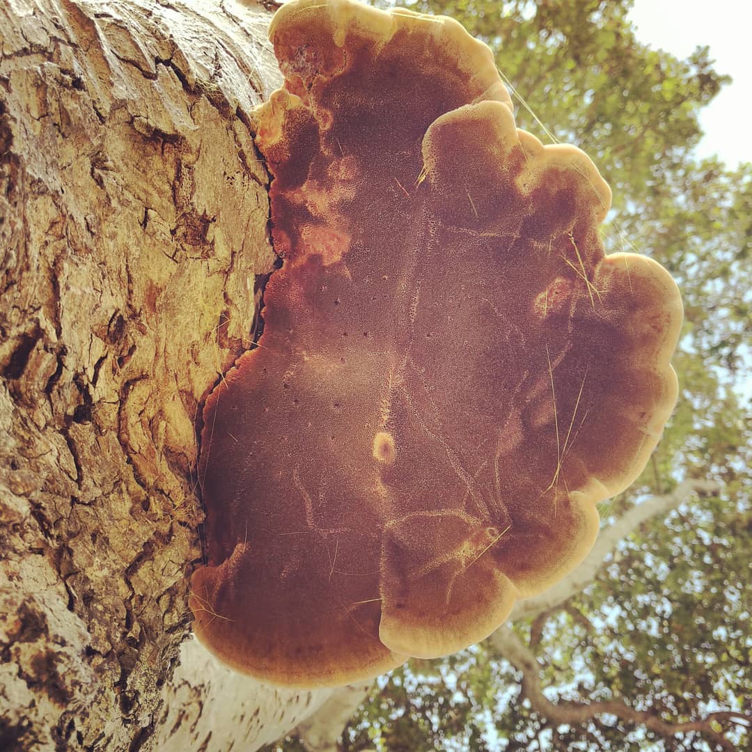
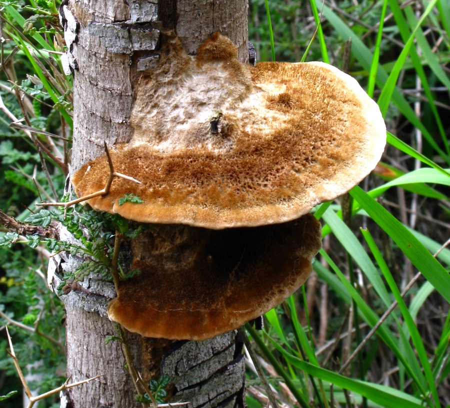
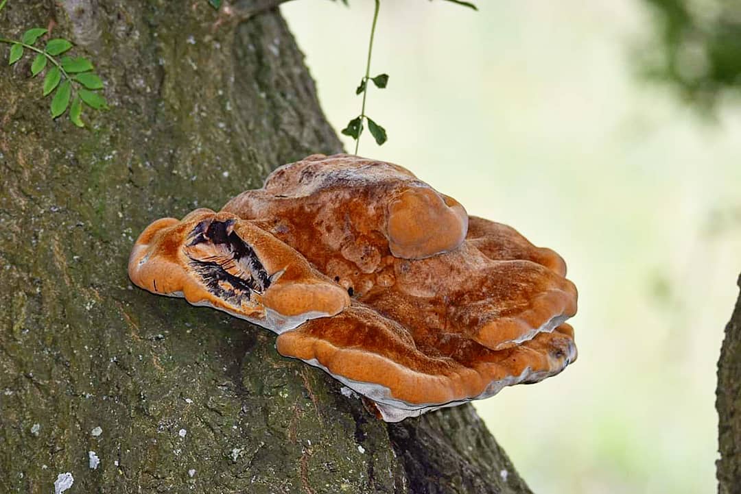
The bristly-haired tinder fungus has fruiting bodies in the form of large fan-shaped outgrowths, 35 cm in size, with an extension at the base. At the initial stage, the tinder fungus has a light yellow color. In the process of development, the fungus acquires a dark brown color and the so-called bristles. As a rule, tinder fungi appear on trees at the end of May.
The fruit body has a very dense and firm structure. The surface is coated with dense fibers. It contains a large number of concentric grooves. The surface of the mushroom is flat, slightly concave. The pores are round or angular. By themselves, they are very small. Depending on the weather conditions, the pulp can be soft and spongy and well saturated with water when the weather is damp, and dry and fragile when there has been no rain for a long time. Spores have a smooth structure and thick walls, similar in appearance to wide ellipses, and the size can reach 7-12 × 6-9 microns.
Indications and restrictions for use
Healing wood sponge is recommended for various diseases. The list of indications for internal use is quite long and includes diseases of many body systems. These include:
- oncology;
- diseases of the heart and blood vessels;
- leukopenia;
- viral diseases;
- inflammatory processes;
- diabetes mellitus and other disorders of the endocrine system;
- damage to the mucous membrane of the duodenum;
- gastritis;
- the transferred operation;
- insomnia, depression and other nervous disorders;
- obesity;
- cramping;
- tachycardia and hypertension.
Chaga birch mushroom In addition, the mushroom is used for general strengthening of the body and increasing immunity, to stimulate brain activity. Chaga is also used externally to treat diseases such as:
- dermatosis;
- rash;
- colds on the lips;
- keratinization of the integumentary epithelium;
- abscesses;
- burns;
- frostbite;
- scuffs, abrasions and wounds;
- acne;
- peeling of the skin;
- insect bites.
The use of the mushroom for the treatment of diseases In dentistry, it is used as an anti-inflammatory and analgesic agent for:
- deep damage to the periodontal tissue;
- inflammatory disease of periodontal tissues;
- damage to the oral mucosa;
- toothache.
However, it is not always possible to use tinder fungus for medicinal purposes. Contraindications to the use of the mushroom are:
- taking penicillin drugs;
- intravenous administration of glucose solution;
- insufficient bowel movements;
- inflammatory disease of the colon mucosa;
- shigellosis;
- pregnancy;
- lactation period;
- individual intolerance.
Advice!
During the treatment period, it is recommended to replace meat with vegetable and dairy sources of protein. You should not eat canned and smoked foods, spicy foods. In addition, you should give up bad habits such as smoking and alcohol abuse.
Thuja diseases
Young shoots and leaves, which turn yellow or acquire a reddish-brown color, are affected. Sporulation of the pathogen is formed on rounded dying areas of the bark and leaves, which looks like crowded black-brown rounded tubercles with a diameter of up to 0.3 mm protruding from the breaks of the integumentary tissues.
Pestalocyopsis necrosis (causative agent - fungus Pestalotiopsis funerea)
Pestalocyopsis necrosis
Young shoots and leaves, which acquire a light brown or reddish brown color, are affected. Leaf damage begins from the top of the shoots, spreading downward. On dying and dead leaves and bark, sporulation of the fungus is formed in the form of a few scattered black rounded tubercles with a diameter of up to 0.2 mm protruding from cracks in the integumentary tissues. Mature spores emerge on the surface of affected leaves and shoots in the form of dark brown, almost black drops and thin cords, which are a characteristic sign of the disease.
Phomopsis necrosis (causative agent - fungus Phomopsis juniperovora)
Affected shoots and leaves turn brown. On the dead bark and in the axils of the leaves, sporulation of the pathogen is formed in the form of black rounded tubercles. Mature spores emerge on the surface of the affected organs in the form of light droplets or strands.
Cytospore necrosis, or osteoporosis (caused by fungi from the genus Cytospora)
Mostly trunks and branches are affected, less often - leaves, which acquire a brown color. The disease is detected by the sporulation of pathogens, which look like numerous, very small conical tubercles with dark tops. Mature spores come to the surface in the form of well-visible golden-yellow, orange or reddish drops, thin flagella and spirals. (Read more about the disease in one of our articles).
Diplodious necrosis, or diplodiosis (causative agent - the fungus Diplodia thujae)
The bark of trunks, branches and leaves is affected. The color of the bark hardly changes, and the leaves turn brown or reddish-brown. In the dead areas, sporulation of the pathogen is formed, which looks like numerous scattered black rounded tubercles with a diameter of up to 0.5 mm.
Brown shute (causative agent - mushroom Herpotrichia juniperi)
The pathogen develops in winter under the snow, therefore, the leaves of only that part of the crown that is in the snow cover zone in winter are affected. After the snow melts, the infected leaves are covered with dense dark brown mycelium (mycelium), which, as it were, glues the affected shoots. On the mycelium, the fruiting bodies of the fungus are formed in the form of black spherical small tubercles, poorly distinguishable on the brown mycelium.Over time, the mycelium is destroyed, and dirty brown scraps remain on the affected leaves.
Mature thuja trees can become infected with wood-destroying fungi, of which root rot pathogens are the most dangerous: autumn honey fungus (Armillaria mellea), root sponge (Heterobasidion annosum), flat tinder fungus (Ganoderma lipsiense), Schweinitz tinder fungus (Phaeolus schweinitzii).
Honey fungus on the trunk Fruit bodies of the root sponge Fruit body of the tinder fungus
All these diseases occur, as a rule, against the background of a preliminary weakening of thuja caused by various unfavorable factors (weather conditions, imbalance of nutrients in the soil, damage by pests, etc.).
Tinder fungus harsh
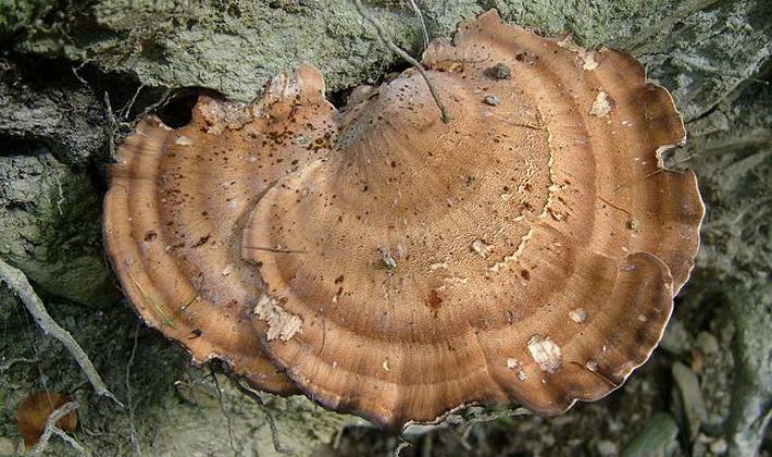
Stiff-haired tinder fungus (lat.Trametes hirsuta).
Fruit body:
Fruit bodies are annual, overwintering, usually in the form of semicircular or kidney-fan-shaped sessile caps, less often prostrate-bent or rosette-shaped, adherent to the tree with a small base, 4-12 cm in diameter, as a rule, flat, thin (0.3-1, 5 cm), single or collected in tiled groups. The surface is coarse-wooled or bristly, concentrically zonal-furrowed (sparse zones), from whitish, ash-gray to yellowish or brownish gray, sometimes with a greenish tint from algae settling on it. Growing bundles of relatively long hairs (up to 4–5 mm), vertical, coarse, hard, more or less brittle, gray. The edge is sharp, thin, sometimes wavy or lobed.
Tubular layer:
The hymenophore is tubular, off-white in youth, then grayish and finally gray (sometimes with a brownish tint). The tubules are single-layered, 1–4 mm long. The pores are thick-walled, rounded, equal in size, with solid margins, 2–4 x 1 mm.
Spore powder is white. Spores are colorless, non-amyloid, without a drop, smooth, narrow-cylindrical, slightly curved, with one pointed end, 5 - 8 x 1.5 - 2.5 microns.
Pulp:
The tissue is thin, leathery, flexible, usually indistinctly zoned, white or very pale colored, fibrous-cotton-like when broken, becomes hard and dense with age and when it dries. The taste is slightly bitter. Young specimens may have a slight aniseed odor.
Habitat:
On stumps, branches, dead, dead and dying trunks of deciduous trees (bird cherry, alder, birch, willow, oak, rowan, beech, hornbeam, aspen, poplar, apple, pear, etc.). It grows both in shady forests (especially often in bird cherry thickets), and in forest glades, clearings. It can grow in wooden buildings and on fences located near the edge of the forest. Occurs throughout the warm period, in mild climates all year round. Frequent species, distributed in the Northern temperate zone.
Notes:
Trametes pubescent Trametes pubescens is distinguished by thinner fruiting bodies with a light-colored, non-zonal, fine-hairy surface of the cap and a yellowish hymenophore with thin-walled unequal pores. Cerenna monochromatic Cerrena unicolor has a tissue with a dark line and hymenophores with unequal, devaliform, lenzites-like, sometimes irpex-like pores at the end, and less elongated spores. Lenzites birch Lenzites betulina is distinguished by a hymenophore: in young specimens it is labyrinth-like, in adults it is lamellar.
Composition and medicinal properties
Chaga contains elements that, in combination, provide a healing effect. The list of biologically active substances contained in a birch mushroom consists of:
- flavonoids;
- alkaloids;
- tannins;
- groups of organic acids.
Each of the elements in the composition of chaga has an individual effect of a therapeutic nature:
- organic acids control and normalize the acid-base balance of the human body;
- flavonoids have anti-inflammatory, antispasmodic, diuretic and choleretic effect;
- phytoncides provide antimicrobial effect;
- alkaloids have a beneficial effect on the heart muscle;
- tannins strengthen and restore the mucous membrane and skin (used for bleeding and inflammation);
- melanin stimulates metabolic processes and restores the body.
Additionally, chaga contains minerals and trace elements. Of these, the most beneficial to human health are:
- magnesium - effective for diseases of bones, joints, teeth, heart, gastrointestinal tract, nerve tissues;
- potassium - helps to treat diseases of the blood, heart, kidneys, and has an antitoxic effect;
- iron - normalizes blood formation and tissue respiration, liver and spleen function, prevents anemia;
- manganese - strengthens bone tissue, improves the absorption of vitamins, relieves inflammation;
- copper - has a beneficial effect on hemoglobin, skin, hair, cellular respiration, oxygen supply, the construction of bone tissue, the work of the nervous system.
Also, the composition of chaga is filled with zinc, cobalt, nickel, silver and aluminum. Most of the elements contained in birch mushrooms are beneficial to human health. Therefore, chaga is used not only in medicine, but also in cosmetology.
The use of chaga in medicine
In official medicine, the following chaga medicines are used:
- Chaga, crushed dried raw materials - for the preparation of decoctions, infusions and tinctures.
- Alcohol tincture - as a general tonic for the treatment of depression, juvenile acne, spasms of internal organs.
- Broth - for diabetes mellitus to lower sugar levels, for chronic gastritis, intestinal atony. For a long time, the broth is used as an auxiliary therapy in the treatment of cancer.
- Infusion - for gastric diseases, to restore the body after infectious diseases and operations. Outwardly, the infusion is used for rinsing the mouth with tonsillitis, stomatitis, periodontal disease. Inhalations with infusion help to eliminate hoarseness, relieve inflammation in the airways.
- Befungin is a chaga extract combined with cobalt chloride. It is used to treat psoriasis, gastritis, gastrointestinal dyskinesia, intestinal atony, tumor pathologies.
What is a tinder fungus
This is a large group of saprophytes that belong to the section Basidiomycetes. They parasitize on wood, leading to the death of the plant. But, unlike chaga, tinder fungi sometimes grow in soil.
You can find them in park areas, in pastures, along the roadside.
In contrast to the canted inonotus, tinder fungi have prostrate, sedentary bodies in the form of a semicircle, a flattened sponge or a large hoof. The consistency of their pulp is hard, woody, corky or spongy.
The stem of the fruiting body is often absent.
But there are known species in which this part of the sporocarp did not atrophy.
This group of basidiomycetes is characterized by a tubular hymenophore, but some representatives of the species are distinguished by a spongy structure. The shape and weight of different types of tinder mushrooms is strikingly different. Some specimens can be up to 1.5 m in size and weigh up to 2-3 kg.
Autumn honey agaric
Autumn honey fungus (Armillaria mellea) is the causative agent of white sapwood (peripheral) rot of roots and trunks. Currently, many researchers consider it not as a separate species of fungi, but as a complex of closely related species, differing in areas, morphological and biological features.
Many conifers and deciduous species, as well as fruit crops are affected. Often, honey fungus occurs as a saprotroph on dry, deadwood and stumps, but under certain conditions it turns into a parasitic way of life and infects living trees and shrubs, causing them to dry out.
Mushroom on the trunk
Autumn honey agaric is widespread in different categories of forest and urban plantings.
- External signs of the disease in conifers are manifested in the thinning of the crown, yellow-green, yellow-brown or brown color of the needles, the presence of cracks and resinification in the butt of the trunks.
- When deciduous species are affected, the crowns of diseased trees become openwork due to crushing of leaf blades. Premature fall of foliage is often observed, cracks form in the butt of the trunks, from which a mucous liquid sometimes flows out.
The characteristic modifications of the mycelium (mycelium) in the form of films and rhizomorphs and fruiting bodies (basidiomas) are reliable signs of autumn honey fungus.
White fan-shaped films form under the bark of thick roots and trunks, covering a significant part of their surface. At first they are thin, but over time they thicken, turn yellow, partially split and transform into rhizomorphs.
- Rhizomorphs develop under the bark of roots and trunks and on the surface of the roots.
- Subcrustal rhizomorphs have the appearance of dark brown, flat, branching cords.
- The outer rhizomorphs are dark brown, almost black, rounded in cross section, similar to the roots of higher plants. They spread up to 30 m and infect healthy roots. Outside rhizomorphs can also move from infected roots to healthy ones through the litter.
Rhizomorphs of the mushroom
The most active development and distribution of rhizomorphs occurs at high humidity and temperatures from +17 ° C to 25 ° C.
Fruiting bodies (basidiomas) of autumn honeydew have the appearance of annual caps on a central leg. Caps are convex or flat, often with a tubercle in the center, yellowish brown, grayish brown, covered with darker scales. The hymenophore is lamellar, white. Stem up to 10-15 cm long, slightly thickened at the base, light brown, fine-scaled, with a white fluffy ring under the cap. Fruiting bodies are formed in groups on old stumps, deadwood and dead wood trunks. In very rare cases, basidiomas can be found on the roots and at the base of the trunks of affected living trees.
Honey mushroom films
The basidiospores formed on the hymenophore plates are spread by air currents, rainwater, animals and infect stumps and roots. The most active formation of basidiospores, their scattering and infection occurs at the end of summer - in autumn in humid warm weather.
Autumn honey fungus affects woody plants, as a rule, against the background of their preliminary weakening caused by various unfavorable factors (weather conditions, damage by other diseases, damage by pests, industrial air and soil pollution, etc.).
Taxonomy
Synonyms:
- Cycloporellus Murrill, 1907
- Flaviporellus Murrill 1905
- Inoderma P. Karst., 1879 non (Ach.) Gray, 1821 nec Berk., 1881
- Inodermus Quél., 1886
- Inonotopsis Parmasto, 1973
- Phaeoporus J. Schröt., 1888
- Polystictoides Lázaro Ibiza, 1917
- Xanthoporia Murrill, 1916
List of species
- Inonotus adnatus Ryvarden, 2002
- Inonotus afromontanus Ryvarden, 1999
- Inonotus albertinii (Lloyd) P.K. Buchanan & Ryvarden, 1988
- Inonotus andersonii (Ellis & Everh.) Černý, 1963
- Inonotus australiensis Ryvarden, 2005
- Inonotus austropusillus Ryvarden, 2005
- Inonotus baumii (Pilát) T. Wagner & M. Fisch., 2002
- Inonotus boninensis T. Hatt. & Ryvarden, 1993
- Inonotus chihshanyenus T.T. Chang & W.N. Chou, 1998
- Inonotus chilanshanus T.T. Chang & W.N. Chou, 2000
- Inonotus clemensiae Murrill, 1908
- Inonotus costaricensis Ryvarden, 2002
- Inonotus cuticularis (Bull.) P. Karst., 1879
- Inonotus dentatus Ryvarden, 2002
- Inonotus dentiporus Ryvarden, 2002
- Inonotus diverticuloseta Pegler, 1967
- Inonotus dryadeus (Pers.) Murrill, 1908
- Inonotus duostratosus (Lloyd) P.K. Buchanan & Ryvarden, 1988
- Inonotus euphoriae (Pat.) Ryvarden, 2005
- Inonotus farlowii (Lloyd) Gilb., 1976
- Inonotus fimbriatus L.D. Gómez & Ryvarden, 1985
- Inonotus flavidus (Berk.) Ryvarden, 1984
- Inonotus fulvomelleus Murrill, 1908
- Inonotus glomeratus (Peck) Murrill, 1920
- Inonotus gracilis Ryvarden, 2005
- Inonotus hamusetulus Ryvarden, 1984
- Inonotus hemmesii Gilb. & Ryvarden, 2002
- Inonotus hispidus (Bull.) P. Karst., 1879 - Inonotus, or bristly-haired tinder fungus
- Inonotus iodinus (Mont.) G. Cunn., 1948
- Inonotus japonicus Ryvarden, 2005
- Inonotus juniperinus Murrill, 1908
- Inonotus leporinus (Fr.) P. Karst., 1882
- Inonotus levis P. Karst., 1887
- Inonotus lloydii (Cleland) P.K. Buchanan & Ryvarden, 1993
- Inonotus lonicericola (Parmasto) Y.C. Dai, 2010
- Inonotus luteoumbrinus (Romell) Ryvarden, 2005
- Inonotus magnisetus Y.C. Dai, 2010
- Inonotus marginatus Ryvarden, 2002
- Inonotus micantissimus (Rick) Rajchenb., 1987
- Inonotus microsporus Ryvarden, 1999
- Inonotus mikadoi (Lloyd) Gilb. & Ryvarden, 2000
- Inonotus munzii (Lloyd) Gilb., 1969
- Inonotus navisporus Ryvarden, 2005
- Inonotus neotropicus Ryvarden, 2002
- Inonotus nidus-pici Pilát ex Pilát, 1953
- Inonotus nodulosus (Fr.) P. Karst., 1882
- Inonotus nothofagi G. Cunn., 1948
- Inonotus novoguineensis Ryvarden, 2005
- Inonotus obliquus (Ach.ex Pers.) Pilát, 1942 - Chaga
- Inonotus ochroporus (Van der Byl) Pegler, 1964
- Inonotus pachyphloeus (Pat.) T. Wagner & M. Fisch., 2002
- Inonotus pacificus Ryvarden, 2005
- Inonotus palmicola Ryvarden, 1999
- Inonotus papyrinus Ryvarden, 2005
- Inonotus patouillardii (Rick) Imazeki, 1943
- Inonotus pegleri Ryvarden, 1975
- Inonotus peristrophidis S. Ahmad, 1972
- Inonotus pertenuis Murrill, 1908
- Inonotus plorans (Pat.) Bondartsev & Singer, 1941
- Inonotus porrectus Murrill, 1915
- Inonotus pruinosus Bondartsev, 1962
- Inonotus pseudoglomeratus Ryvarden, 2002
- Inonotus pseudoradiatus (Pat.) Ryvarden, 1983
- Inonotus pusillus Murrill, 1904
- Inonotus quercustris M. Blackw. & Gilb., 1985
- Inonotus radiatus (Sowerby) P. Karst., 1881 - Radiant inonotus
- Inonotus rheades (Pers.) Bondartsev & Singer, 1941 - Fox Polypore
- Inonotus rickii (Pat.) D.A. Reid, 1957
- Inonotus rodwayi D.A. Reid, 1957
- Inonotus setulosocroceus (Cleland & Rodway) P.K. Buchanan & Ryvarden, 1993
- Inonotus shoreae (Wakef.) Ryvarden, 2005
- Inonotus sideroides (Lév.) Ryvarden, 2005
- Inonotus splitgerberi (Mont.) Ryvarden, 1972
- Inonotus subiculosus (Peck) J. Erikss. & Å. Strid, 1969
- Inonotus tabacinus (Mont.) G. Cunn., 1948
- Inonotus triqueter (Fr.) P. Karst., 1882
- Inonotus tropicalis (M.J. Larsen & Lombard) T. Wagner & M. Fisch., 2002
- Inonotus ulmicola Corfixen, 1990
- Inonotus ungulatus Ryvarden, 2005
- Inonotus vaninii (Ljub.) T. Wagner & M. Fisch., 2002
- Inonotus venezuelicus Ryvarden, 1987
- Inonotus weirianus (Bres.) T. Wagner & M. Fisch., 2002
- Inonotus weirii (Murrill) Kotl. & Pouzar, 1970
- Inonotus xanthoporus Ryvarden, 1999
External signs of raw materials
text_fieldstext_fieldsarrow_upward
External signs
Rice. 11.17.2. Oblique tinder
Whole raw material Pieces of various shapes up to 10 cm in size. The outer layer of the build-up is black, strongly cracking, the inner layer is dark or brown-brown with small yellow veins, the number of which increases towards the inner side. The tissue of the fungus is dense and hard. There is no smell. Bitter taste.
Chopped raw material Pieces of raw material passing through a sieve with holes with a diameter of 7 mm. Color dark brown. There is no smell. Bitter taste.
Impurities
Sometimes collectors mistakenly collect other fungi parasitizing on birch.
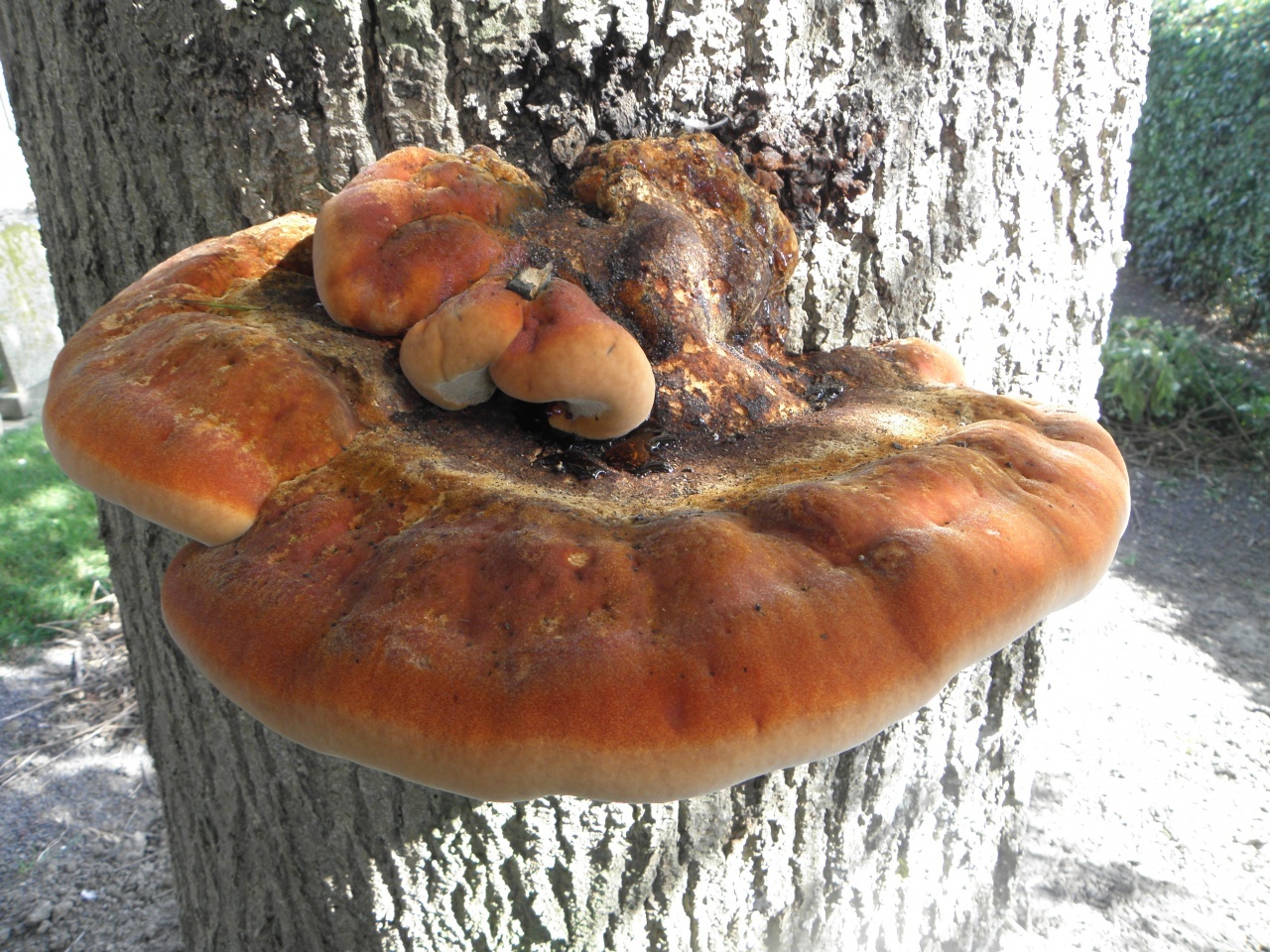
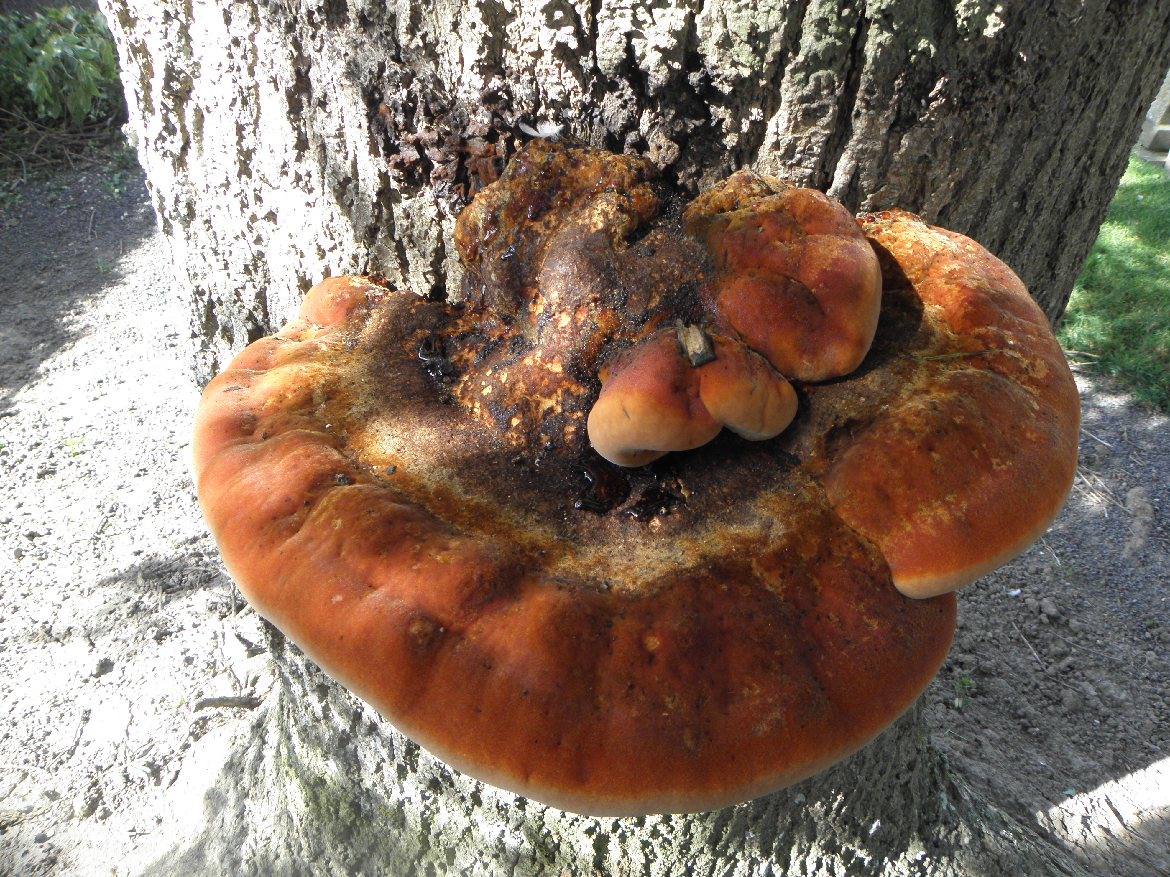
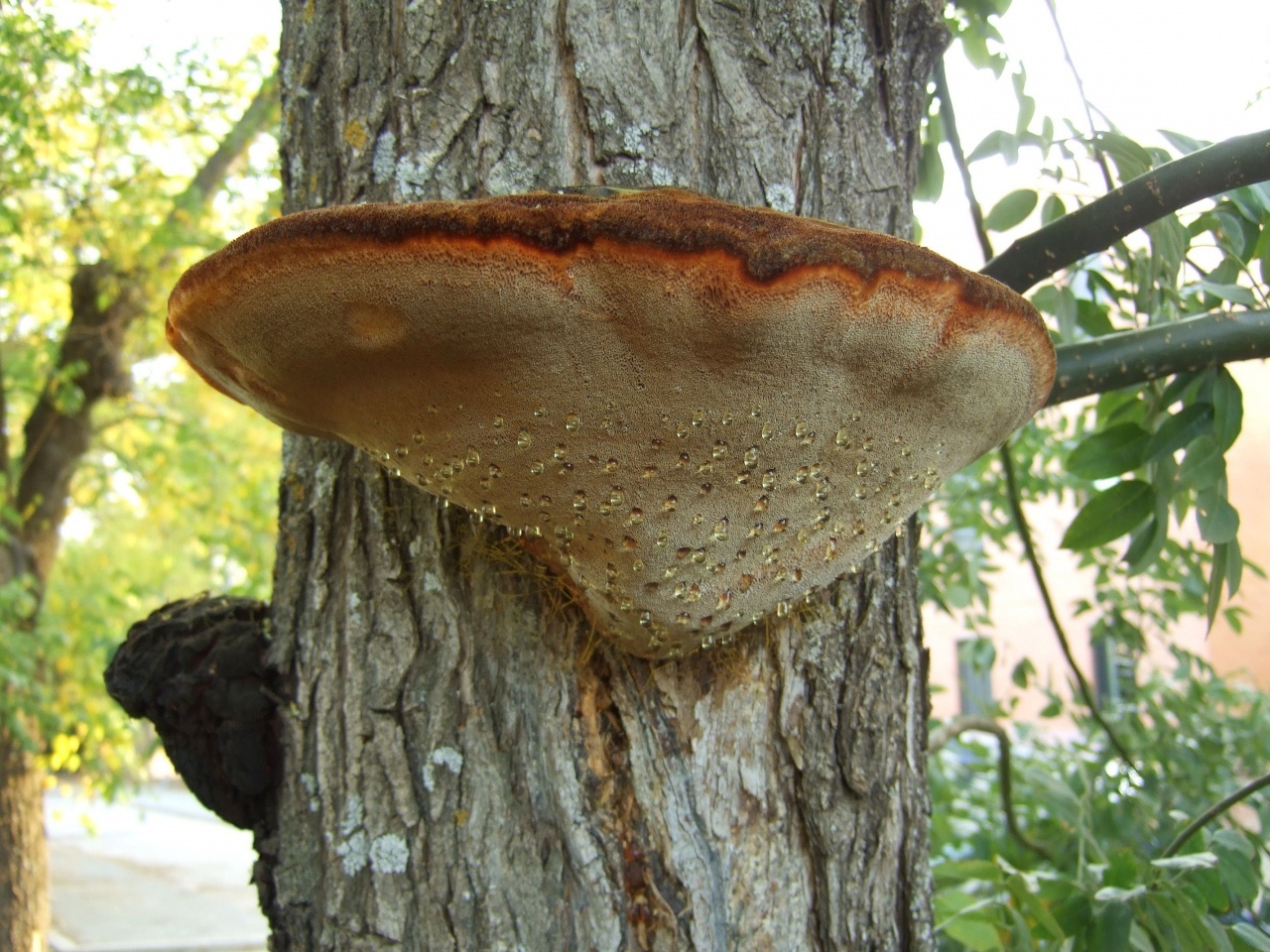
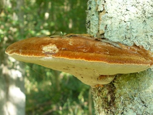
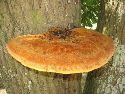
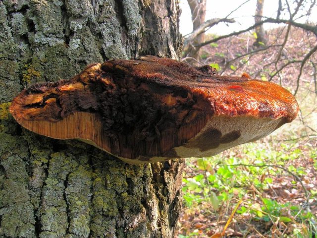
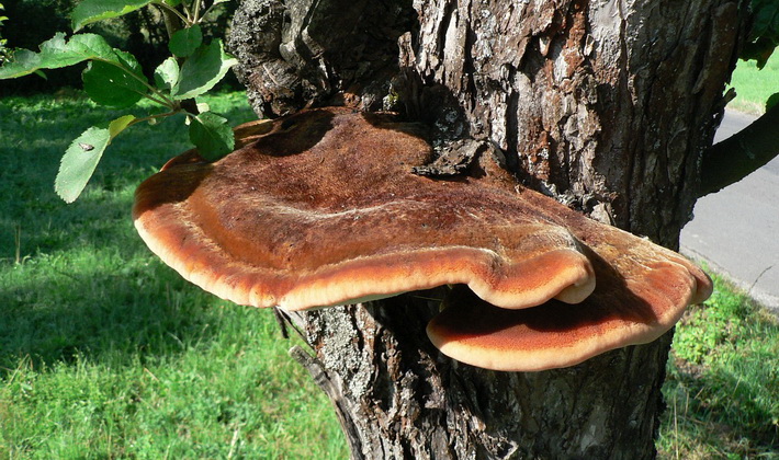
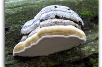
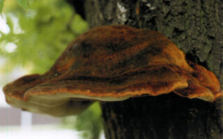
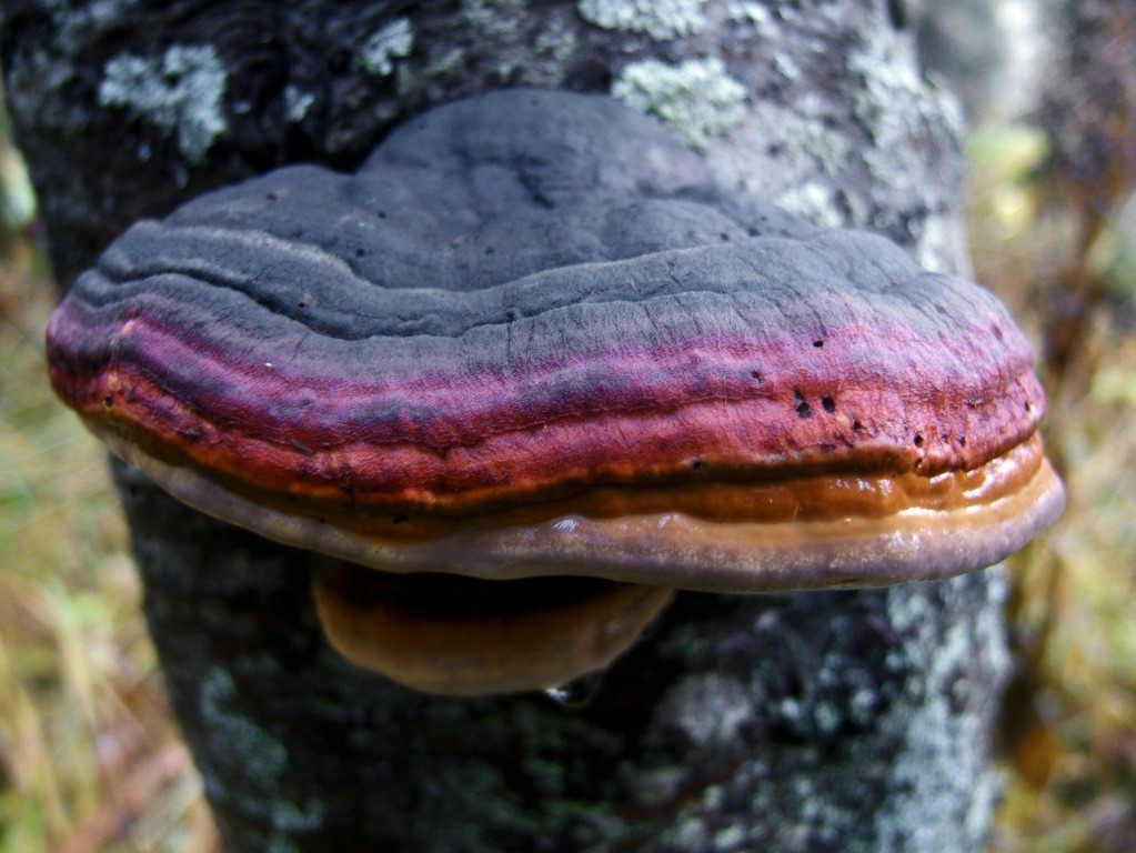
Most often, real and false tinder fungus come across. Both fungi develop a fruiting body, which has a hoof-like shape, convex above, flat below with a velvety surface (hymenial layer).
Distinctive features of a birch fungus from similar species
|
Mushroom name |
Diagnostic signs |
|
|
fruiting body shape |
surface |
|
| Chaga | Oval or rounded | Dark brown, pitted or cracked, with many small bumps and fissures |
| False tinder fungus | Hoof-shaped, facing flat side down (top convex) or having the appearance of a hat | Velvety, with concentric circles, hard, with a grayish-black or black-brown crust |
| Real tinder | Hoof-shaped, semicircular in outline, flat on the underside, with a wide base | Smooth, with concentric grooves, hard, with a grayish or brownish crust. Wavy layers are noticeable |
Destruction of wood
Drying of trees in the foci of the root sponge
Rotting wood is the process of its decomposition and destruction by wood-destroying fungi using a specific set of enzymes (substances that convert complex organic compounds into water-soluble, easily digestible by the fungus).
The nature of the destruction depends on the type of fungus and its set of enzymes, the degree and sequence of its destruction of cell walls, changes in the chemical composition of wood pulp and its physical properties. There are destructive and corrosive types of decay.
In the destructive type, decomposition of cellulose and hemicellulose occurs, which are the main constituent part of the cell walls and provide mechanical strength and elasticity of wood tissues. The cell membranes are destroyed evenly, gradually, without the formation of large holes in them. The fungus affects the entire mass of wood. As a result, the entire volume of the wood decreases, numerous cracks appear in it. Subsequently, the wood disintegrates into prismatic pieces, becomes brittle, and is easily ground into powder. The color of the wood is also gradually changing. At first, it becomes reddish, later turns brown, and in the final stage it acquires a dark brown color. Rot of this type is caused by the Swiss tinder fungus, sulfur-yellow tinder fungus, bordered tinder fungus, larch sponge and birch sponge, etc.
With a corrosive type of decay, lignin (an organic compound that causes lignification of the cell walls) and partially the cellulose complex are destroyed. Some fungi simultaneously decompose lignin and cellulose, destroying groups of cells in separate places. In the affected wood, cavities appear in the form of pits and cells, filled with white undecomposed cellulose. White spots of cellulose on a brown wood background create a variegated rot color (variegated corrosive rot). Variegated corrosive rot is caused by a root sponge, a pine sponge and a sponge sponge, spruce butt tinder fungus and oak tree tinder fungus.
Corrosive rot from root sponge
Other types of fungi initially completely decompose lignin, and then cellulose and other polysaccharides are gradually destroyed. At the same time, in the final stage of decay, the affected wood brightens evenly or in stripes, acquires a white, light yellow or "marble" color (white corrosive rot).White corrosive rot is caused by autumn honey fungus, real tinder fungus, false tinder fungus, flat tinder fungus.
Root rot is the most dangerous for growing trees, causing them to weaken, dry out, and reduce resistance to wind.
In the case of a corrosive type of decay, not all of the wood mass is decomposed: individual groups of destroyed cells alternate with untouched areas of wood. Therefore, at different stages of wood destruction, rot acquires a pitted, pitted-fibrous, fibrous structure. The wood splits into fibers, crumbles, retains its viscosity, and its volume does not decrease.
Fruit bodies of the root sponge
Tinder fungus sulfur-yellow and birch
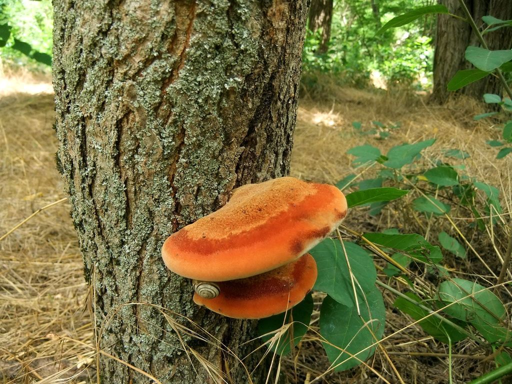
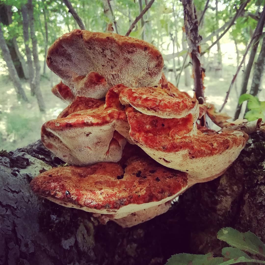
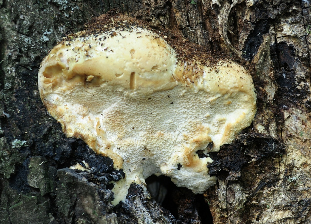
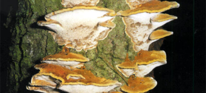
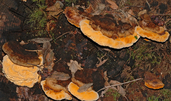
Tinder fungus is sulfur-yellow. Fruit bodies 6-30 cm wide, first thick pineal, then semicircular or fan-shaped, tiled, adherent sideways, fleshy, juicy, later dry and brittle, sulfur-yellow in youth, later yellow-orange, and finally ocher. The light yellow or pale ocher surface of the cap is covered with brown scales. On the underside of the cap, angular and elongated, rather large pores of the hymenophore. The tubular layer below is represented by small sulfur-yellow, later yellow-ocher pores. The pulp of young sulfur-yellow tinder fungi is soft, juicy, brittle, white. The smell is weak, in old tinder fungi it is unpleasant, in young mushrooms it is lemon, the taste is sour.
It grows on the trunks and stumps of deciduous and coniferous trees. Bears fruit in spring.
Birch polypore. Fruit bodies 4–20 cm in diameter, convex to almost flattened, 2–6 cm thick. The crust of young fruiting bodies is whitish, later gray, yellowish or pale brown. The fabric is white. The layer of tubules is separated from the tissue. The surface of the hymenophore is white, subsequently slightly brown. The spore powder of the birch fungus is white. Spores 4.5-6 x 1.2-1.5 µm, cylindrical, smooth, colorless.
Growth. Grows on dead, rarely living, birches.
Fruiting. July to December.
This is one of those tinder fungi that cause a yellowish-brown or reddish-brown rot of a destructive type, which develops intensively. The wood struck by this tinder fungus quickly collapses and becomes rotten. When infected, rotting first develops in the bark and sapwood, and from there quickly penetrates to the center of the trunk; mushroom caps develop in the last stages of wood decay. In the initial stages of the lesion on cross cuts, rot appears in the form of a complete or incomplete peripheral ring of wood with a reddish tint, gradually turning into reddish brown or yellowish brown. Subsequently, cracks in the radial and tangential directions are noticed on the affected wood.


