Damage
Due to the fact that the structure of the ethmoid bone is porous, the segment is very susceptible to injury. Fractures often occur in an accident, in a fall, in a fight, an antero-ascending blow to the nose. Fragments of bone can freely move through the ethmoid plate, in fact, into the cranial cavity. This can provoke cerebrospinal fluid (CSF) in the nasal area. The resulting message of the cranial and nasal cavities provokes severe, difficult to eliminate infections of the central nervous system. The ethmoid bone has a close connection with the olfactory nerve. If the element is damaged, sensitivity to odors may deteriorate or completely disappear.
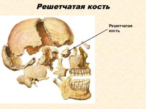
Ethmoid bone
(os ethmoidak)
, unpaired, consists of 2 plates: lattice, located horizontally, and perpendicular (Fig. 1, 2), as well as a lattice labyrinth. A labyrinth is a paired formation, represented by a complex of air cells adjacent to a lattice plate on each side. The cells communicate with each other and with the nasal cavity.
Rice. 1.
Ethmoid bone:
a - topography of the ethmoid bone;
b - top and back view: 1 - perpendicular plate; 2 - the wings of the cock's comb; 3 - front cells of the lattice labyrinth; 4 - back and middle cells of the lattice labyrinth; 5 - orbital plate; 6 - lattice plate; 7 - cock's comb;
c - bottom view: 1 - perpendicular plate; 2 - upper nasal concha; 3 - lattice plate; 4 - middle turbinate; 5 - hooked process; 6 - back cells of the lattice labyrinth; 7 - front cells of the lattice labyrinth;
d - view from the lateral surface: 1 - cockscomb; 2 - the wings of the cock's comb; 3 - front cells of the lattice labyrinth; 4 - perpendicular plate; 5 - hooked process; 6 - middle turbinate; 7 - orbital plate
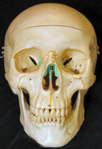
Rice. 2.
The participation of the ethmoid bone in the formation of the walls of the cranial cavity, eye sockets and nasal cavity
1 - ethmoid bone; 2 - cranial cavity; 3 - eye socket; 4 - maxillary sinus; 5 - nasal cavity
The ethmoid bone is located in the ethmoid notch of the frontal bone. The ethmoid plate of the ethmoid bone is part of the cerebral section of the skull. The rest of the parts take part in the formation of the bony walls of the nasal cavity and the medial walls of the orbits.
Lattice plate
(lamina cribrosa)
connected in front and on the sides with the frontal bone, behind - with the anterior edge of the sphenoid bone. The plate is penetrated by many small lattice holes (foramina cribrosa)
for the branches of the olfactory nerves. The cockscomb (crista galli) extends upward from the lattice plate along the midline
, to which the anterior end of the cerebral crescent is attached. In front of this ridge lies a paired process - the wing of the cock's comb (ala cristae galli)
.
Perpendicular plate
(lamina perpendicularis)
of an irregular hexagonal shape, descends downward, forming the anterior part of the bony septum of the nose.
Lattice maze cells
(labirintus ethmoidale)
divided into three groups, not sharply delimited from each other: anterior
, average
and back
... From the lateral side, they are covered with a very thin bony orbital plate (lamina orbitalis)
facing the free surface into the cavity of the orbit. On the inner side, only a small part of the lattice cells is covered with bone plates. Most of them remain open and are covered by adjacent bones: frontal, lacrimal, wedge-shaped, palatine and upper jaw.
The medial surface of the labyrinth limits the upper part of the nasal cavity and is equipped with 2 thin bone plates facing the nasal cavity - the upper
and middle turbinates (conchae nasales superior et media)
as well as the hooked process (processus uncinatus)
... There is a gap between the shells - the upper nasal passage (meatus nasi superior)
... Above and behind the superior concha, the highest nasal concha (concha nasalis suprema) is sometimes found
... Ethmoid shells have different shapes and sizes, determining different depths and lengths of the corresponding passages of the nasal cavity.
Ethmoid bone ossification begins with ethmoid plasty and labyrinth at 5-6 months of intrauterine development. At the end of the 1st year of life, centers of ossification appear at the base of the cock's comb and in the perpendicular plate. The fusion of parts of the bone occurs in the 5-6th year.
Human anatomy S.S. Mikhailov, A.V. Chukbar, A.G. Tsybulkin
Techniques for correcting the joints (sutures) of the ethmoid bone
Correction of the wedge-lattice seam
IPP - lying on your back
IPV - at the head of the patient, from the side
Cephalic hand 1 and 3 fingers capture the large wings of the sphenoid bone and the outer pillars of the frontal bone
Caudal hand - 1 toe on the nasion (optional), 2 toes on the cruciform suture, 3 toes in front of the cruciforms (behind the teeth).
- synchronization with LDM
- in the phase of cranial expiration, we induce the ethmoid bone into extension due to the cephalic effect on the cruciate suture
- in the phase of cranial expiration, follow the movement of the sphenoid bone
- save the obtained parameters, wait for the point of balanced tissue tension (still point) and tissue relaxation
- we induce the sphenoid, frontal, ethmoid bones into flexion

Correction of the frontal lattice suture
IPP - lying on your back
IPV - at the patient's head
Stage 1
Arms positioning as with frontal lift
Aggravation technique
- in the phase of cranial expiration, to transfer the ethmoid bone into flexion, there is a slight medial effect on the external pillars of the frontal bone and its transfer to extension (movement in the ventral and caudal directions)
- at the phase of cranial inspiration, we hold the obtained parameters
- we carry out the action for 1-2 cycles of the LDM until a feeling of resistance appears on the outer pillars of the frontal bone and the beginning of tissue movement at the level of the lattice notch
“Stage 2
Setting hands like frontal spread
Direct technique
- at the phase of cranial inspiration, we lift the ventrally and translate laterally the external pillars of the pubic bone
- we apply medial pressure on the glabella to open the lattice notch
- within several LHD cycles we win in parameters
- expect an increase in the mobility of the KSM
IPP - lying on your back
IPV - at the head of the patient, on the side of him
Placement of hands - the cephalic hand grasps the frontal bone and large wings of the sphenoid bone, the caudal hand with 2 and 3 fingers contacts the anterior-inner surface of the upper jaws, 1 finger is on the nasion
- synchronization with LDM
- in the phase of cranial inspiration, induction of the frontal bone into flexion
- at the next phase of cranial inhalation, we part 2 and 3 fingers of the caudal hand laterally, 1 finger presses on the nasion
- win in parameters for several LHD cycles
- the result of performing the technique is an increase in freedom and amplitude in the area of the nasion
Examples of osteopathic correction techniques
- Drainage of venous sinuses
- SBS decompression techniques
- Frontal lift
- Parietal lift
- Compression technique of the fourth ventricle of the brain (CV4, compression of ventricle four)
- Correction of the fronto-parietal suture
- Correction of the occipital-parietal suture
- Correction of the parietal scaly suture
- Correction of the parieto-mastoid suture
- Correction of temporo-occipital sutures
- Bregma correction
- Temporal bone correction
- Ethmoid bone testing and correction
- Dysfunction of the pelvic bones
Symptoms of ethmoiditis
Symptoms of acute primary ethmoiditis. Acute ethmoiditis often manifests itself as follows:
- Frequent headaches;
- Painful sensations in the area of the inner edge of the orbit;
- Difficulty nasal breathing;
- Hyposmia (decreased sense of smell), or anosmia (complete absence of smell);
- Deterioration of the general condition;
- Subfebrile temperature (up to 38 degrees) in the first 48 hours of the disease and its sharp increase (up to 40 degrees) in the following days;
- Discharge from the nose (discharge is abundant, colorless and odorless) in the first 2-3 days;
- Discharge from the nasal cavity with an admixture of pus (after 3 days of illness) with an admixture of mucus;
- Redness and swelling of the inner corner of the orbit (only in children);
- Phenomena of toxicosis, which are growing rapidly (nausea, vomiting);

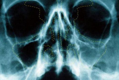
X-ray with ethmoiditis
Secondary acute ethmoiditis has more striking manifestations in the form of:
- septic processes (there are metastatic purulent foci);
- tension and infiltration of the eyelids;
- the appearance of a bluish skin tone of the eyelids;
- immobility of the eyeball;
- a decrease in the diameter of the nasal passages and a complication of respiratory function.
Nasal conch
From the medial side, the cells are covered with curved thin bone plates. They represent the middle and upper turbinates of the nose. The lower edge of each hangs loosely into the gap. It runs between the perpendicular plate and the labyrinth. The upper portion of each shell is attached to the medial surface of the labyrinth openings. Above, respectively, the upper shell is attached, just below it and slightly anteriorly is the middle one. In some cases, a third element is also found. It is called the "highest shell" and is rather weakly expressed. The nasal passage lies between the middle and upper shells. It is represented by a narrow gap. The middle course is located under the curved side of the corresponding turbinate. It is limited from below by the upper portion of the inferior nasal concha. On its posterior edge, there is a hook-shaped process curved downwards. It articulates on the skull with the ethmoid process extending from the inferior shell. Behind this formation, a large bubble protrudes into the middle course. This is one of the largest cavities that the ethmoid bone includes. Behind and above, between the large vesicle and the uncinate process, a gap is visible in front and below. It has the shape of a funnel. Through this gap, the frontal sinus and the middle nasal passage are communicated. This is the normal ethmoid anatomy.


Anatomy and biological defense
 Skull bones
The structure of the lattice labyrinth is a system of tunnels and cavities that is quite complex at first glance. But upon closer examination, one can easily distinguish three main groups of it - anterior, middle and posterior.
Hard tissues of the middle surface of the labyrinth (medial) limit the upper part of the nasal cavity. There are also two thin bone plates facing the nasal cavity, which form two turbinates - the upper and middle. Depending on their size and shape, the appearance of the human nose changes. Between these shells is the main nasal passage.
It is important to mention the downward-curved process located at the top of the lower portion of the nasal concha. Behind it is a very voluminous anatomical cavity, which connects the frontal sinus and the middle nasal passage, with the help of a clearly visible funnel-shaped gap
Danger of injury
The porous structure of the ethmoid bone due to the anatomical features of such a structure has an extremely low resistance to mechanical damage. As a result, a fight, a fall, and any accidents associated with an antero-incoming blow to the nasal region can cause this bone to break. Its main danger is that, due to its porous structure, ethmoid bone easily breaks into separate fragments, each of which can damage soft tissues. Often, the result of a blow to the nose is liquorrhea.
So you shouldn't underestimate this unusual bone, which is located in a very hidden place for a reason. Ethmoid bone is indeed the most fragile of the bones of the skull. It is worth remembering that injuries that damage the bone in question are extremely dangerous.
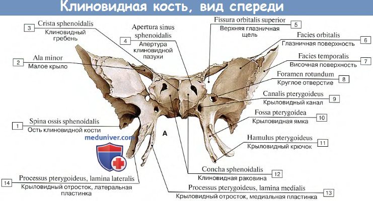
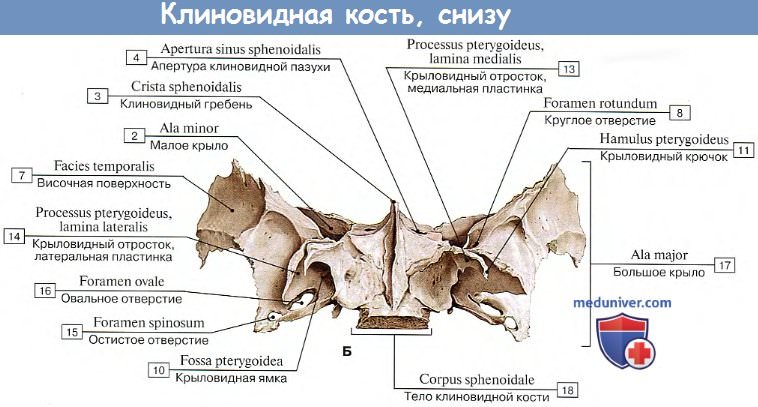
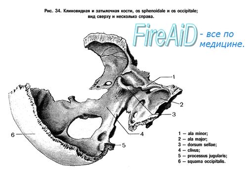
Anatomy: Sphenoid bone
Sphenoid bone, os sphenoidale, unpaired, resembles a flying insect, which explains the name of its parts (wings, pterygoid processes).
The sphenoid bone is the product of the fusion of several bones that independently exist in animals, therefore it develops as a mixed bone from several paired and unpaired ossification points, forming 3 parts by the time of birth, which in turn fuse into a single bone by the end of the first year of life.
The following parts are distinguished in it:
1) body, corpus (in animals - unpaired basisphenoid and presphenoid);
2) big wings, alae majores (in animals - paired alisphenoid);
3) small wings, alae minores (in animals - paired orbitosphenoid);
4) pterygoid processes, processus pterygoidei (its medial plate is a former paired pterygoid, develops on the basis of connective tissue, while all other parts of the bone arise on the basis of cartilage).
Body, corpus, on its upper surface has a depression along the middle line - Turkish saddle, sella turcica, at the bottom of which there is a hole for pituitary gland, fossa hypophysialis.
In front of her is eminence, tuberculum sellae, along which it passes transversely sulcus chiasmdtis for cross (chiasma) optic nerves; at the ends sulcus chiasmatis the visual channels are visible, canales opticithrough which the optic nerves pass from the cavity of the eye sockets into the cavity of the skull. Behind the Turkish saddle is limited by a bone plate, saddle back, dorsum sellae.
On the lateral surface of the body there is a curved sleep sulcus, sulcus caroticus, trace of the internal carotid artery.
On the front surface of the body, which is part of the back wall of the nasal cavity, ridge visible, crista sphenoidalisbelow, entering between the opener wings. Crista sphenoidalis connects in front with the perpendicular plate of the ethmoid bone. Irregular shapes are visible on the sides of the ridge holes, aperturae sinus sphenoidalisleading to the airway sinus sphenoidalis, which is placed in the body of the sphenoid bone and is divided septum, septum sinuum sphenoidalium, into two halves. Through these openings, the sinus communicates with the nasal cavity.


In a newborn, the sinus is very small and begins to grow rapidly only around the 7th year of life.
Small wings, alae minores, are two flat triangular plates, which with two roots extend forward and laterally from the anterosuperior edge of the body of the sphenoid bone; between the roots of the small wings are the mentioned visual channels, canales optici. Between the small and large wings is superior orbital fissure, fissura orbitalis superiorleading from the cranial cavity to the orbital cavity.
Big wings, alae majores, depart from the lateral surfaces of the body laterally and upward. Near the body, behind fissura orbitalis superior there is round hole, foramen rotundumleading anteriorly to the pterygo-palatine fossa, due to the passage of the second branch trigeminal nerve, n. trigemini... Posteriorly, a large wing in the form of an acute angle juts out between the scales and the pyramid of the temporal bone. Near him there is spinous hole, foramen spinosumthrough which passes a. meningea media.
Much more can be seen in front of it. oval hole, foramen ovalethrough which the third branch of item trigemini passes.
Large wings have four surfaces: cerebral, facies cerebralis, orbital, facies orbitalis, temporal, facies temporalis, and maxillary, facies maxillaris... Surface names indicate the areas of the skull where they face. The temporal surface is divided into the temporal and pterygoid parts by infratemporal crest, crista infratemporalis.
Pterygoid processes processus pterygoidei depart from the junction of the large wings with the body of the sphenoid bone vertically downward. Their base is permeated with a sagittal canal, canalis pterygoideus, - the place of passage of the same nerve and blood vessels. The anterior opening of the canal opens into the pterygo-palatine fossa.
Each process consists of two plates - lamina medialis and lamina lateralis, between which the back is formed fossa, fossa pterygoidea.
The medial plate is bent at the bottom crochet, hamulus pterygoideus, through which the tendon begins on this plate is thrown m. tensor veli palatini (one of the muscles of the soft palate).


- We also recommend "Anatomy: Temporal bone"
Ethmoid bone biomechanics
Ethmoid bone is an unpaired symmetrical midline bone. The biomechanics of this bone is determined by its structure.The central part (horizontal plate) defines the movement as the bones of the midline (flexion, extension). The lateral masses determine the external and internal rotation.
Axes of movement:
- Horizontal axis (flexion, extension) - passes transversely through the body of the bone at the level of the anterior-inferior part of the cock's crest
- Vertical axis - defines the movement of the external and internal rotation
Phase of LDM flexion
The ethmoid bone experiences the traction of the cockscomb from the side of the stretching sickle ligament, while:
- the apex of the cock's crest moves cephalic and dorsally
- the back of the lattice plate descends
- the back of the perpendicular plate descends
- lateral masses move laterally (external rotation)
Phase of extension of LDM
As a result of weakening the tension of the sickle, the ethmoid bone returns back, while:
- the apex of the cock's comb moves ventrally
- the back of the lattice plate rises
- the back of the perpendicular plate rises
- lateral masses move medially
Structural features
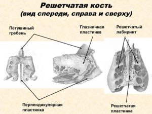 Ethmoid bone in three projections
The anatomy divides the complex of tissues called ethmoid bone into three main sectors. The first is the ethmoid plate, which is located at the top of the bone. As you know, a feature of the structure of the ethmoid plate is the presence of a large number of holes for the fibers of the olfactory nerve. In addition, there is a small growth on it, called the "cock's comb". In front of this growth, there is a blind hole that includes a part of the frontal bone.
The perpendicular plate is the second element of the aggregate of the considered complex of bone tissue. It is located in the sagittal plane and is an element that forms the upper part of the nasal septum.
Trauma
All joints represent a complex system, the violation of which threatens with serious consequences for human health. The area of the nose most often suffers from falls or blows with the face part, since the cartilage protrudes significantly above the surface of the face. When this area is damaged, the entire nasal mucosa is injured, subcutaneous emphysema appears. After the injury, the face swells. The swelling spreads to the neck area. If the ethmoid artery is damaged, there is a risk of bleeding into the orbit. This can affect vision.
This form of injury is positioned with a fracture of the skull. This type of injury occurs in almost half of traumatic brain injuries. The most common causes of damage are:
- falling on the face from a height;
- hitting your face on the steering wheel during an accident;
- inflicting a direct blow on the face of the skull in the area of the nose.
The main risk of injury lies in the possible consequences. For example, due to the ingress of microorganisms into the cranium, through their introduction by air, infection develops. If, upon receipt of damage, first aid was not provided or treatment was not carried out, an inflammatory process occurs, which turns into a purulent form. This form of a pathological condition is extremely dangerous for human life and is fatal. The inflammation itself, even not in a purulent form, is dangerous, since complications arise with the development of such a process.
In some cases, injury is accompanied by paralysis of the nerves of the surface of the face, less often - fatal. Statistics show that a small percentage of such trauma is accompanied by paralysis of the whole body for an indefinite period of time with different outcomes. This phenomenon suggests that when this part of the face was damaged, the nerve fibers associated with the spinal nerve were affected.
The reason that the bone is injured much more easily than others is its porous structure.With a splinter fracture, there is a high risk of the fragment entering the cranial box, which can cause cerebrospinal fluid to leak. This pathology is called liquorrhea, and has dangerous consequences, since the receipt of useful substances by the brain is disrupted. Sometimes the pneumatization of the cells is disrupted or polyps appear.
If the plate that protects the eye socket is damaged, the eye "rolls out", for example, when sneezing. Trauma, accompanied by breaks in the nerve fibers of the sense of smell, can partially, and sometimes completely deprive the person of the ability to smell.
Such an injury should be taken as seriously as possible, since the bone is located close to the brain, and if severely damaged, it can touch it. In addition to unpleasant sensations, damage threatens consequences that are dangerous for general health and condition.
Treatment
When the ethmoid labyrinth is inflamed, conservative treatment is most often prescribed to the patient. At the initial stage, vasoconstrictor drugs are recommended to the patient, which help to reduce swelling and increase the outflow of separated mucus. Also, the patient is prescribed antibiotics and pain relievers. After a few days, physiotherapy procedures begin.
If a patient is diagnosed with a chronic form, then the treatment is no different from the acute one. In the remission stage, surgery may be recommended.

Brain department
The vault contains flat bones. These include the scales of the temporal and occipital, as well as the frontal and parietal elements. Flat bones consist of plates of compact substance (internal and external), between which lies a cancellous bone structure (diploe). The connection of the roof elements is carried out by means of seams. At the base of the skull - the lower part - is the foramen magnum. It connects the cavity to the spinal canal. There are also holes for nerves and blood vessels. The pyramids of the temporal elements act as the lateral bones of the base. They contain the departments of the organs of balance and hearing. Allocate the inner and outer sides of the base of the skull. The first is divided into rear, middle and front center pits. They contain different parts of the brain. In the central part, in the middle pit, there is a Turkish saddle. The pituitary gland lies in it. On the outside of the base, two condyles lie to the sides of the foramen magnum. They are involved in the formation of the atlantooccipital joint.


Classification of ethmoiditis
- Acute emoiditis (primary, secondary);
- Chronic ethmoiditis;
- Polypoid emoiditis (a separate type, or as a subspecies of chronic ethmoiditis)
Acute ethmoiditis. The main cause of acute ethmoiditis is the aggravation of rhinitis, flu and other inflammatory diseases. Very often, inflammation of the paranasal sinuses develops into a lesion of the ethmoid labyrinths. During the acute phase of inflammation, the anterior cells are involved in the process (if the person suffered from frontal sinusitis or sinusitis). The posterior cells of the ethmoid bone are affected by inflammation of the sphenoid sinuses. The acute inflammatory process proceeds extremely quickly, therefore it is strictly forbidden to delay treatment. The mucous membrane swells diffusely, which leads to narrowing and then closing of the excretory ducts of the ethmoid cells. If the inflammation spreads to the bone, then it manifests itself in the form of the formation of fistulous and abscessing passages.
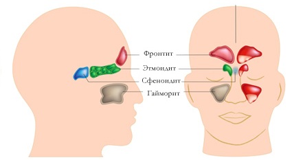
Varieties of sinusitis
Prophylaxis
Since the inflammation of the mucous membrane of the ethmoid labyrinth can be caused by various microorganisms, there are no specific preventive measures.
In order to avoid provoking factors as much as possible, it is necessary to try to prevent the occurrence of diseases that can provoke inflammation.In addition, you need to monitor your immunity, give preference to a healthy and balanced diet, give up bad habits, and take immunomodulatory agents in the fall and spring. Also, if signs appear that may indicate the development of an inflammatory process, immediately contact a medical institution and strictly follow all the recommendations of your attending physician.
Ethmoid plate of ethmoid bone
This element is the top of the segment. It is located in the ethmoid notch in the frontal bone. The plate is involved in the formation of the bottom in the anterior cranial fossa. The entire surface of the element is occupied by holes. In appearance, it resembles a sieve, hence, in fact, its name. Through these holes, the olfactory nerves (the first pair of cranial) run into the cranial cavity. Cockscomb is present along the median line above the plate. In the anterior direction, it continues with a paired process - a wing. These parts, together with the frontal bone, which lies in front, delimit the blind foramen. In some way, the perpendicular surface acts as a continuation of the ridge. It has an irregular pentagonal shape. It is directed downward towards the nasal cavity. In this zone, the plate, located vertically, participates in the formation of the upper region of the septum.
Therapeutic activities
Treatment usually requires special care. After the diagnosis is made, the doctor prescribes antibacterial drugs to avoid blood poisoning. To stop bleeding, an appropriate hemostatic drug is prescribed. If there are various unpleasant symptoms, for example, severe headaches have appeared, drugs are prescribed that will correspond to the symptoms.
In addition, sedatives, various analgesics and others are often required. Without fail, the patient is injected with a serum against tetanus infection.
In this way, the doctor helps the patient to overcome the inflammatory process, to avoid the development of infection in the body, since during the general therapy, the body is significantly weakened and becomes susceptible to pathogenic bacteria.
If the injury was serious, there was a displacement, or the fracture was shrapnel, an operation is required. This procedure is rarely performed, but in case of blockage of the sinuses of the nose or severe deformation, it is imperative to return a person to a normal lifestyle.

For the period of therapy, it is necessary to abandon any heavy physical and mental stress. Nutrition should be balanced, with a predominance of foods that contain calcium in the diet. After the nasal mucosa is restored, specialized nasal drops with a vasoconstrictor effect may be required.
Trauma diagnosis
Diagnostics can only be carried out by a doctor in a hospital setting. The doctor conducts a brief assessment of the expressed symptoms, if necessary, palpation of the damaged area is performed. Information about the injury received is compulsorily collected, first of all, about the prescription of the receipt. The doctor then learns how the injury was sustained, subsequent conditions, such as bouts of nausea, vomiting, or dizziness with loss of consciousness. In addition, information on previous facial injuries will be required.
After palpation of the swelling, the doctor gives a general assessment of the patient's condition and sends it to laboratory tests. Laboratory tests include a study of urine, blood, and nasal discharge.
An obligatory procedure is an electrocardiogram. Then the doctor decides which examination method is most appropriate. First of all, an X-ray procedure is prescribed in order to identify the fracture.Thanks to computed tomography, it is possible to identify the presence of displacement of debris or an area of tissue rupture. Ultrasound examination determines how much damage is. In more rare cases, an endoscopy or lumbar puncture is required.

Clinical signs
Inflammation of the cells of the ethmoid labyrinth in an acute form is characterized by pronounced symptoms. Signs of pathology can occur suddenly and manifest themselves with strong intensity. Most often, patients complain of intense headaches, which are manifested mainly in the area of the eye socket and nose. When the head is tilted, the pain sensations increase. In addition, a person may be disturbed by shortness of breath, as well as nasal discharge of a mucous or mucopurulent consistency. In some situations, the patient may show symptoms of intoxication, weakness, fatigue, and body temperature rise.
As for the course of the disease in childhood, compared with adults, the condition is much more severe. The manifestation of pathology begins with an increase in body temperature. The child becomes restless, completely refuses to eat. If you do not seek qualified medical help in a timely manner, then the child may develop signs of neurotoxicosis, as well as dehydration.


When the first signs of inflammation appear, you should immediately seek qualified medical help. Otherwise, ethmoiditis can turn into a chronic stage, the treatment of which is very difficult.
Damage symptoms
Fractures of any area are always accompanied by the appearance of swelling, soreness or bruising
But it is important to remember that such external signs do not in all cases indicate a fracture of the ethmoid bone. In certain cases, it can be a bruise or a crack, which is less dangerous for a person's life.
Therefore, it is necessary to know what other signs indicate a serious injury to the ethmoid bone.
Damage to the nasal region is characterized by the following features:
- visible deformation of the site;
- nosebleed;
- damage to the skin on the bridge of the nose or around the nasal region;
- any uncharacteristic discharge from the nose.

If the bone has been damaged, then the above symptoms appear immediately after injury. Any injury requires attention and referral to specialists.
Appointment
The sinuses of the ethmoid labyrinth are complex. They still remain the subject of scientific research. This is explained by the fact that scientific knowledge, unfortunately, is not enough to fully determine the origin and stage-by-stage development.
The ethmoid labyrinth of the nose performs the following functions:
- protective - since there is air in the cavity of the sinuses, then upon impact it extinguishes the negative effect on the skull;
- baroreceptor - thanks to this function, signals are transmitted to the body indicating changes in atmospheric pressure;
- moisturizing - provided due to the fact that in the process of air circulation inside the sinuses, it heats up, and then, after contact with the mucous membranes, they are moistened;
- thermal insulation - prevents hypothermia and smoothes out sharp temperature drops during breathing.

Among other things, thanks to the nasal sinuses, the weight of the skull bones is significantly lightened, but the necessary volume is retained.