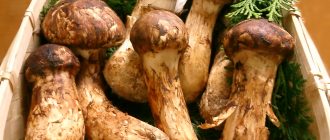
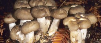
Description of the lingonberry exobasidium
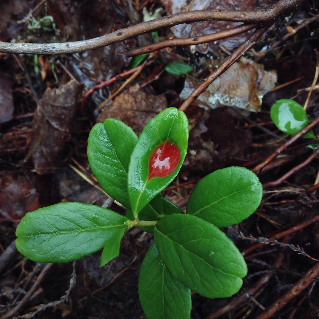
Lingonberry exobasidium does not have a fruiting body. 5-7 days after plant infection, yellow-brown spots form on the upper side of its leaves.

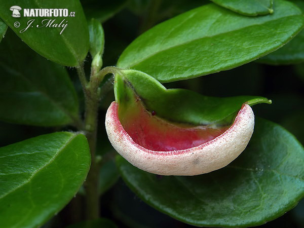
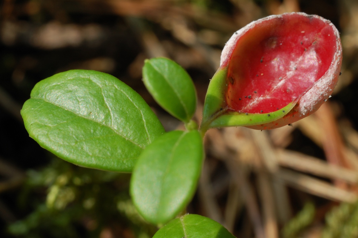
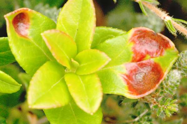
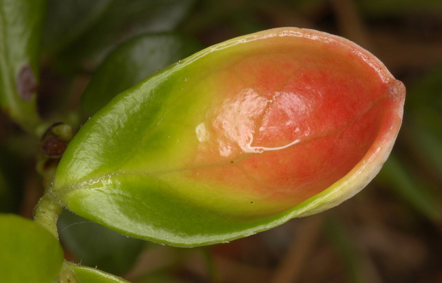
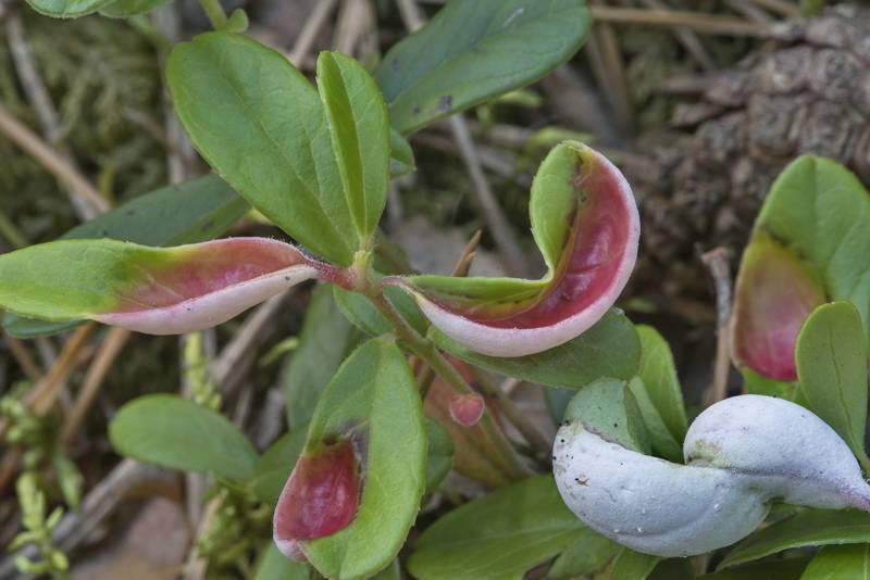
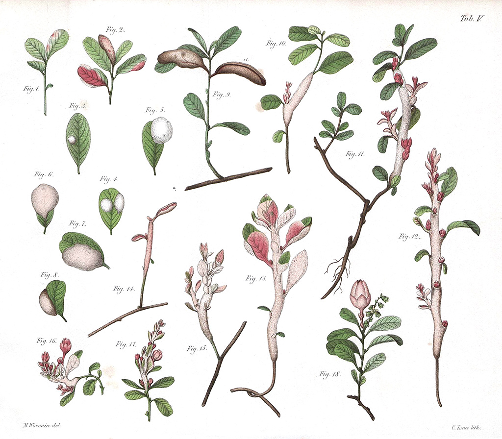
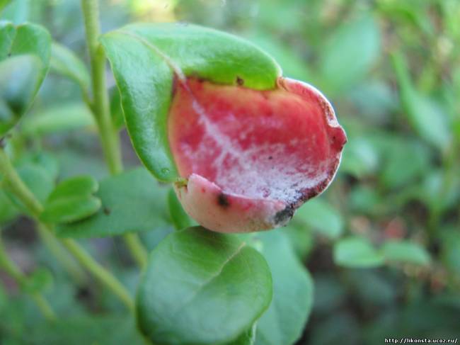
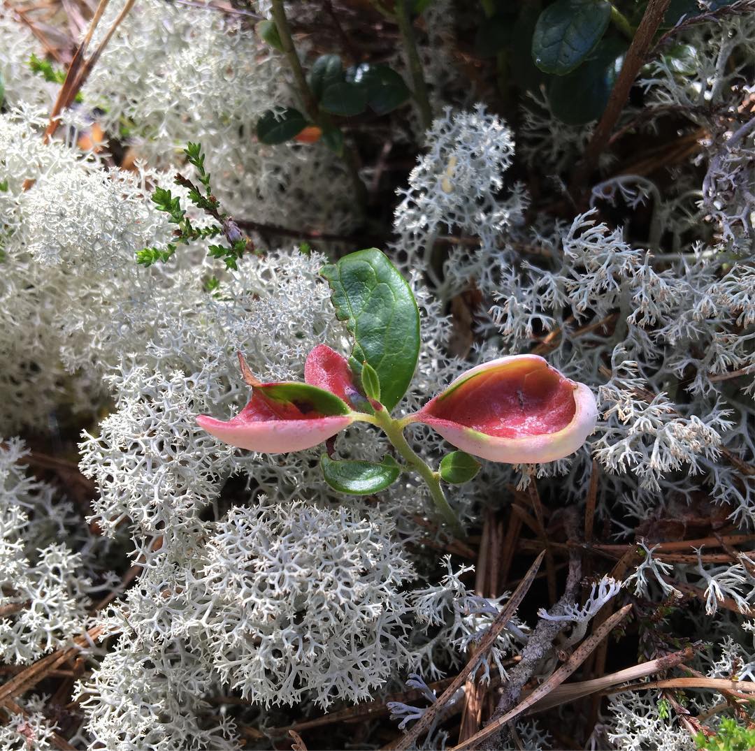
After a week, the spots turn red. The spot can occupy a certain part of the leaf or completely the leaf. The stain is depressed from above, the sheet is deformed. The spot size is 0.5-0.8 centimeters, and its depth is 0.2-0.3 centimeters.
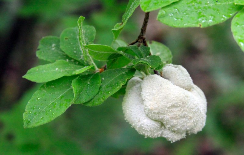
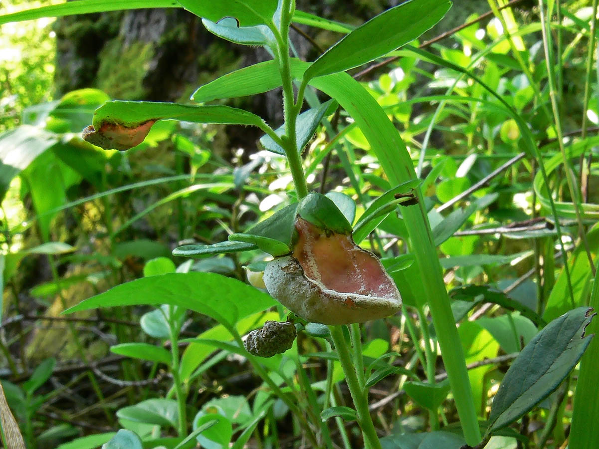
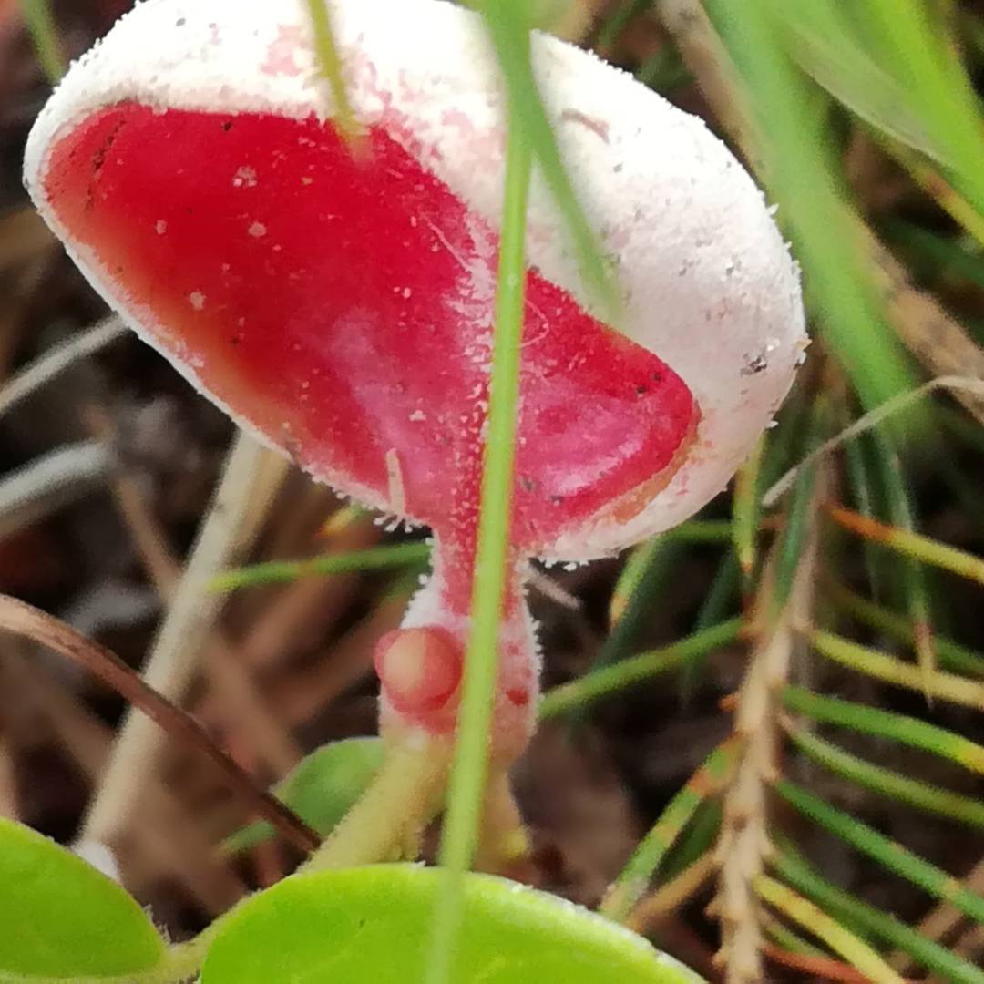
There is a thickened bulge at the bottom of the leaf. This bulge looks like a tumor-like growth. Its diameter is approximately 0.5 centimeters. The surface of the growth is uneven, there is a white bloom on it - these are basidiospores. The pulp is fleshy, dense, white, emits a mild powdery odor.
Distribution of lingonberry exobasidium
Fruiting of lingonberry exobasidium occurs from summer to autumn. These mushrooms settle on young shoots, stems, leaves, and less often on lingonberry flowers. You can meet them in coniferous forests.
This mushroom comes across very often, it grows in almost all taiga forests growing in the northern part and the Arctic.

Influence of lingonberry exobasidium on plants
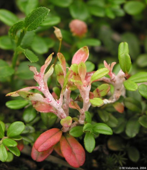
In early or mid-summer, lingonberry leaves begin to deform, sometimes young stems may suffer. The infected areas increase in size. The upper side of the leaves becomes concave, and its color becomes reddish. At the bottom, the infected areas, on the contrary, are convex, and their color is snow-white.
The damaged areas thicken - their thickness is 3-10 times greater in comparison with healthy leaves. Sometimes the stems undergo deformation, they bend and turn white. In rare cases, the fungus infects flowers.
Under a microscope, significant changes in the structure of diseased leaf tissue are clearly visible. The cells are much larger than the usual size, that is, their hypertrophy occurs. Chlorophyll is not produced in the cells of the affected areas. And in the cell sap, a pigment called anthocyanin is formed. It is because of this pigment that the red color of the leaves appears.

Development cycle of lingonberry exobasidium
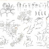
The fungal hyphae are visible between the plant cells. There are more hyphae near the lower surface of the leaves. Thicker hyphae are located between the epidermal cells, here under the cuticle, young basidia are formed.
The cuticle is enlarged, discarded in parts. On each mature basidium, 2-6 fusiform basidiospores are formed. From spores, a delicate white bloom similar to frost is formed on the lower part of the leaf.
When basidiospores enter the water, they quickly become 3-5-cell. On each side of the spore, thin hyphae grow, from which small conidia extend. In turn, they can form blastospores.
When the basidiospores hit the young leaves of the lingonberry, they germinate. The hyphae enter the plants through the stomata of the leaves, and the mycelium forms inside. After 4-5 days, yellowish spots form on the surface of the leaves. A week later, a typical infection pattern appears.

Systematics of the lingonberry exobasidium
Lingonberry exobasidium for mycologists is a reason for disagreement and controversy. Some mycologists claim that exobasidiomycetes are a primitive group that shows that hymenomycetes are descended from parasitic fungi. In this regard, they distinguish these mushrooms in an independent order, placing them ahead of other hymenomycetes.
Opponents consider exobasidiomycetes as a highly specialized group, in the form of a lateral branch of the development of primitive saprotrophic hymenomycetes.
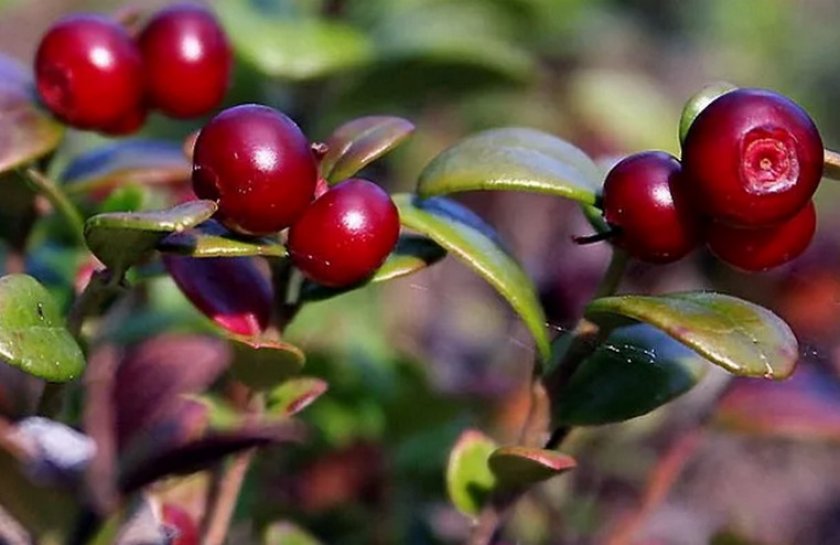
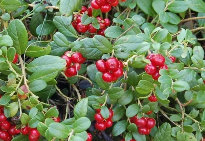
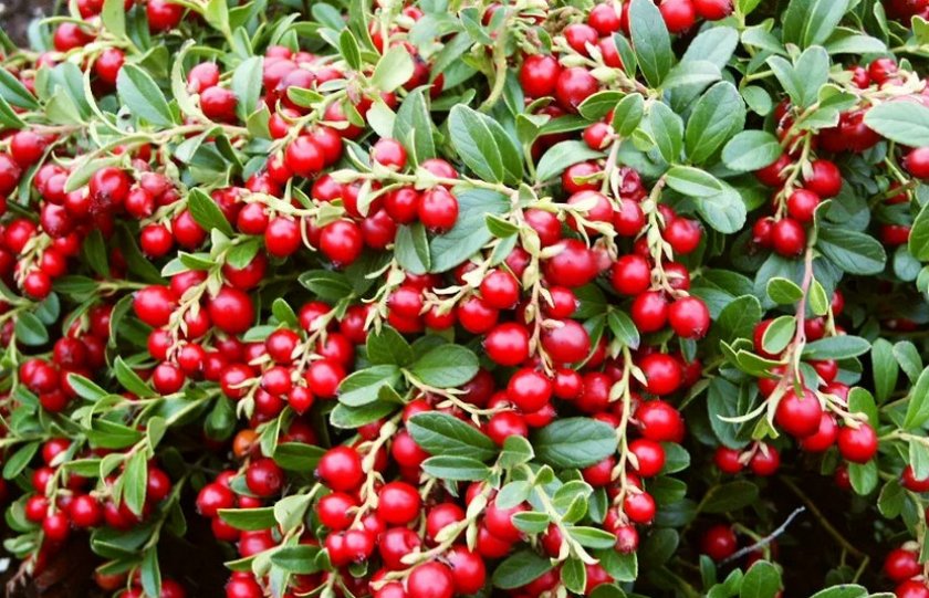
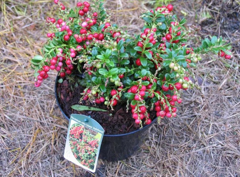
That is, the taxonomy of these fungi is controversial, and the question of the origin of many species of higher fungi remains open. Another aspect raises questions - it is known that lingonberry exobasidium grows not only on lingonberries, but also on bearberry, cranberries, blueberries and other heather plants.Are these mushrooms one species or belong to a whole complex of closely related but independent species?
Definitioner
- Blastospores (Blastospores)
-
A spore formed from a part of a conidiogenic cell that increases in size and is separated by a septum from the conidiogenic cell. The blastic type is divided into two subtypes:
- Holoblastic conidia (blastospores, porospores). Conidia is formed in the form of a kidney on the surface of the conidiogenic cell (sometimes, as it were, "blown out" through the pore), while the conidia membrane is a continuation of the conidiogenic cell membrane. In some fungi, the conidia, after formation, itself turns into a conidiogenic cell and is able to form a new conidia kidney on the surface. With repeated repetition of this process, an acropetal chain of conidia is formed, in which the largest (most mature) is the basal one (eg, Cladosporium, Septonema, Helminthosporium);
- Enteroblastic conidia (phialospores) - the second subtype of blastic spores, unlike holoblastic ones, have their own membrane, which is not a continuation of the conidiogenic cell membrane. In many fungi, the conidiogenic cell (phialida) can repeatedly form conidia, forming basipetal chains, in which the largest (most mature) conidia is the terminal one (for example, in the p. Phialophora, Catenularia).
See Acropetal, Conidia.
- Basidia (Basidia)
-
Lat. Basidia. A specialized structure of sexual reproduction in fungi, inherent only in Basidiomycetes. Basidia are terminal (end) elements of hyphae of various shapes and sizes, on which spores develop exogenously (outside).
Basidia are diverse in structure and method of attachment to hyphae.
According to the position relative to the axis of the hypha, to which they are attached, three types of basidia are distinguished:
Apical basidia are formed from the terminal cell of the hypha and are located parallel to its axis.
Pleurobasidia are formed from lateral processes and are located perpendicular to the axis of the hypha, which continues to grow and can form new processes with basidia.
Subasidia are formed from a lateral process, turned perpendicular to the axis of the hypha, which, after the formation of one basidium, stops its growth.
Based on morphology:
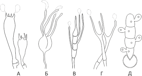
Holobasidia - unicellular basidia, not divided by septa (see Fig. A, D.).
Phragmobasidia are divided by transverse or vertical septa, usually into four cells (see Fig. B, C).
By type of development:
Heterobasidia consists of two parts - hypobasidia and epibasidia developing from it, with or without partitions (see Fig. C, B) (see Fig. D).
Homobasidia is not divided into hypo- and epibasidia and in all cases is considered holobasidia (Fig. A).
Basidia is the place of karyogamy, meiosis and the formation of basidiospores. Homobasidia, as a rule, is not functionally divided, and meiosis follows karyogamy in it. However, basidia can be divided into probasidia - the site of karyogamy and metabasidia - the site of meiosis. Probasidium is often a dormant spore, for example in rust fungi. In such cases, probazidia grows with metabasidia, in which meiosis occurs and on which basidiospores are formed (see Fig. E).
See Karyogamy, Meiosis, Gifa.


