METHODS OF CULTIVATION OF MICROORGANISMS. STUDY OF CULTURAL AND BIOCHEMICAL
PROPERTIES
Cultivation, that is, the cultivation of microorganisms in the laboratory, is used to study their properties and to obtain biomass. Bacteria, fungi, actinomycetes, spirochetes and some protozoa are cultivated on nutrient media. Chlamydia, rickettsia, viruses and some protozoa can reproduce only in the body of an animal or in living cells.
The cultural properties of this type of microorganisms are: 1) the conditions necessary for reproduction, and 2) the nature of growth on nutrient media. Cultural properties are one of the characteristics that are taken into account when identifying (determining the type) of microorganisms.
Culture media
Culture media must meet certain requirements. They must contain all the nutrients necessary for the reproduction of this type of microbes. Some pathogenic microorganisms grow on simple nutrient media, while others need the addition of blood, blood serum, and vitamins for their reproduction.
In culture media, certain conditions must be created by adding sodium chloride or buffer solutions. For most bacteria, a nutrient medium containing 0.5% sodium chloride is favorable. The reaction of the nutrient medium, favorable for most pathogenic bacteria, is slightly alkaline, which corresponds to pH = 7.2-7.4. Vibrio cholerae grows at pH = 7.8-8.5, mushrooms - at pH = 5-5.5. The culture media should be moist, that is, contain a sufficient amount of water, be as transparent and sterile as possible, that is, before sowing, do not contain microbes.
In terms of composition and origin, nutrient media are natural, artificial and synthetic. Natural culture media are natural products such as potatoes and other vegetables. Artificial nutrient media are prepared according to a specific recipe from products with the addition of organic and inorganic compounds. Synthetic media contain certain chemical compounds in known concentrations.
By consistency, nutrient media are liquid, semi-liquid, dense. Agar-agar-polysaccharide isolated from seaweed is usually used as a sealant. Agar-agar is not used by microorganisms as a nutrient; it forms a gel in water that melts at 100 ° C and solidifies at 45 ° C.
To obtain a dense nutrient medium, agar-agar is added at a concentration of 1.5-2%, for semi-liquid - 0.5%.
According to their intended purpose, culture media can be divided into ordinary (simple), special, elective, differential diagnostic.
Conventional (simple) nutrient media are used for the cultivation of most microorganisms, this is mesopatamia broth (MPB), mesopatamia agar (MPA).
Special nutrient media are used for the cultivation of microorganisms that do not grow on simple media. For example, blood agar and sugar broth for streptococcus, serum agar for meningococcus and gonococcus.
Elective culture media are used to isolate one species from a mixture of different bacteria. This type of bacteria grows on this environment faster and better than others, outstripping them in its growth; the growth of other bacteria is delayed on this medium. For example, coagulated serum for diphtheria bacillus, alkaline peptone water for cholera vibrio, bile broth for typhoid bacillus, saline media for staphylococcus.
Differential diagnostic nutrient media are used to distinguish some types of bacteria from others by their enzymatic activity (see the corresponding section).
Cultivation and isolation of pure cultures of aerobic bacteria
For the cultivation of microorganisms, certain conditions are necessary: temperature, aerobic or anaerobic conditions.
The temperature should be optimal for the species. Most pathogenic bacteria thrive at 37 ° C. However, for some species, a lower temperature is optimal, which is associated with the peculiarities of their ecology. So, for the plague bacillus, the natural habitat of which are rodents during hibernation, the optimum temperature is 28 ° C, as for leptospira, for the botulism bacillus - 28 ° C-35 ° C.
In addition to the optimal temperature, for the cultivation of microorganisms, depending on the species, aerobic or anaerobic environment is required.
In order to study the morphology, cultural, biochemical and other properties of microbes, it is necessary to obtain a pure culture. Usually a culture of microbes is called their accumulation on a nutrient medium in the form of turbidity, near-bottom (wall) growth, or a film on the surface of a liquid medium or colonies on a dense medium. A single colony is formed from one microbial cell. A pure culture is a culture of microbes of the same species obtained from a single colony. In laboratories, certain known strains of microbes are used for various studies. A strain is a pure culture of microbes obtained from a specific source, at a specific time, with known properties. As a rule, microbial strains are designated by a specific number. For example, the Staphylococcus aureus 209P strain is used to determine the activity of penicillin.
Isolation of pure cultures of aerobes usually takes three days and is carried out according to the following scheme:
1st day - microscopy of a smear from the test material, stained (usually by Gram) - for a preliminary acquaintance with the microflora, which can be useful in choosing a culture medium for inoculation. Then inoculation of the material onto the surface of the frozen nutrient agar to obtain isolated colonies. Sieving can be performed according to the Drygalsky method into three Petri dishes with a nutrient medium. A drop of material is applied to the first cup and spread with a spatula over the entire cup. Then, with the same spatula, distribute the remaining culture on it on the second cup and in the same way on the third. The largest number of colonies will grow on the first plate, the least on the third. Isolated colonies will grow on one of the plates, depending on how many microbial cells were in the test material.
The same result can be achieved by sieving on one cup. To do this, divide the cup into four sectors. The material under study is inoculated with a bacteriological loop with strokes on the first sector, then, having calcined and cooled the loop, the inoculation is distributed from the first sector to the second and in the same way sequentially into the third and fourth sectors. Isolated colonies are formed from individual microbial cells after 24 h incubation in a thermostat.
2nd day - study of colonies grown on plates, description of them. Colonies can be transparent, translucent or opaque, they have different sizes, rounded regular or irregular outlines, convex or flat shape, smooth or rough surface, smooth or wavy, jagged edges. They can be colorless or white, golden, red, yellow. Based on the study of these characteristics, the grown colonies are divided into groups. Then an isolated colony is selected from the study group, a smear is prepared for microscopic examination in order to check the homogeneity of microbes in the colony. The same colony is inoculated into a test tube with a slant nutrient agar.
3rd day - checking the purity of the culture grown on the agar slant by smear microscopy. With the homogeneity of the studied bacteria, the isolation of a pure culture can be considered complete.
To identify the isolated bacteria, cultural traits are studied, that is, the nature of growth on liquid and solid nutrient media. For example, streptococci on sugar broth form a bottom and parietal sediment, on blood agar - small, pinpoint colonies; cholera vibrio forms a film on the surface of alkaline peptone water, and transparent colonies on alkaline agar; the plague bacillus on nutritive agar forms colonies in the form of "lace handkerchiefs" with a dense center and thin wavy edges, and in a liquid nutrient medium - a film on the surface, and then - threads extending from it in the form of "stalactites".
Cultivation and isolation of pure cultures of anaerobic bacteria
For the cultivation of anaerobes, it is necessary to lower the oxidation-reduction potential of the medium, to create anaerobiosis by removing oxygen by physical, chemical or biological methods.
Physical methods include:
1) mechanical removal of air by means of a pump from anae-rostat, in which dishes with inoculations are placed. At the same time, you can replace the air with an indifferent gas: nitrogen, hydrogen, carbon dioxide.
2) growing in a medium containing reducing substances. Kitta-Tarozzi Wednesday is a sugar broth with pieces of liver or meat. Glucose and pieces of organs have a reducing ability. The medium is poured on top with a layer of vaseline oil to block the access of air oxygen.
3) The simplest, but less reliable method is to grow deep in a tall column of sugar agar.
Chemical methods consist in the fact that dishes with crops of anaerobes are placed in a hermetically sealed desiccator, where chemicals are placed, for example, pyrogallol and alkali, the reaction between which proceeds with the absorption of oxygen.
The biological method is based on the simultaneous cultivation of anaerobes and aerobes on solid nutrient media in Petri dishes, hermetically sealed after inoculation. At first, oxygen is absorbed by the growing aerobes, and then the growth of anaerobes begins.
Isolation of a pure culture of anaerobes begins with the accumulation of anaerobic bacteria by inoculation on Kitta-Tarozzi medium. In the future, isolated colonies are obtained in one of two ways:
1) the material is inoculated by mixing with melted warm sugar agar in glass tubes. After the agar has solidified, isolated colonies grow in its depths, which are removed by cutting the tube and subcultured on Kitt-Tarozzi medium (Weinberg's method);
2) inoculation of the material is carried out on plates with a nutrient medium and incubated in an anaerostat. Isolated colonies grown on a plate are subcultured on Kitt-Tarozzi medium (Zeissler method).
Cultivation of other microorganisms
Cultivation of mycoplasmas
Mycoplasmas are cultured on nutrient media supplemented with serum and carbohydrates. Since mycoplasmas lack a cell wall, they grow only in isotonic or hypertonic environments. On solid nutrient media, for several days, very small colonies are formed, resembling fried eggs - with a convex center and a flat translucent periphery. Mycoplasmas can also be grown in chicken embryos or cell culture.
Cultivation of rickettsia and chlamydia
Rickettsiae and chlamydiae are obligate intracellular parasites. For their cultivation, cell cultures, chicken embryos and animal infection are used.
Mushroom cultivation
For the cultivation of mushrooms, dense and liquid nutrient media are used: most often Sabouraud's medium, as well as media containing beer wort. Mushrooms grow more slowly than bacteria, they form visible growth within a few days. The cultivation temperature is lower than that of bacteria - 22-30 ° C.
Cultivation of spirochetes and protozoa
Among the spirochetes, it is most easy to grow leptospira, for which water mixed with rabbit blood serum can serve as a nutrient medium.Borrelia and treponema are cultivated under anaerobic conditions on more complex nutrient media containing serum, pieces of animal tissue.
Among the protozoa, dysentery amoeba, lamblia, Trichomonas, leishmania, trypanosome, balantidia are cultivated on nutrient media. Toxoplasma is cultivated in chicken embryos and tissue cultures. Cultivation methods for malaria plasmodia are under development.
Methods for studying enzymatic activity (biochemical properties)
In microbiological practice, the study of enzymatic activity is used to identify microorganisms, since each microbial species has a certain set of enzymes.
To determine the proteolytic activity, microbes are inoculated with an injection into a column of gelatin, and after 3-5 days of incubation at room temperature, the character of gelatin liquefaction is noted: in the form of a funnel, a nail, a stocking or in the form of an overturned Christmas tree. Proteolytic activity is also determined by the formation of protein decomposition products: indole, hydrogen sulfide, ammonia. To determine them, microorganisms are inoculated into meat-peptone broth, and indicator papers are placed between the neck of the test tube and a cotton stopper, excluding their contact with the medium. When indole is formed, paper impregnated with a saturated solution of oxalic acid turns pink; in the presence of hydrogen sulfide, paper impregnated with lead acetate turns black; when ammonia is formed, the red litmus paper turns blue.
To determine the saccharolytic properties of microbes, differential diagnostic media are used, such as Giss media, Olkenitsky's medium, Endo's medium, Levin's medium, Ploskirev's medium.
Media Endo, Levin, Ploskirev in Petri dishes are used to differentiate bacteria of the intestinal group by their ability to ferment lactose. These media contain nutrient agar, lactose and an indicator that changes color in an acidic medium - a pH indicator. If bacteria that ferment lactose, such as E. coli, are sown on such an environment, acid is formed as a result of fermentation of lactose, and the indicator will change color in an acidic environment. Therefore, colonies of Escherichia coli on such media will be colored according to the color of the indicator: on Endo's and Ploskirev's medium - in red, on Levin's medium - in black and blue. Colonies of bacteria that do not ferment lactose, such as salmonella and dysentery sticks, will be colorless.
Giss's media (“variegated range” media) are prepared on the basis of peptone water or semi-liquid meat-peptone agar. Contain any one carbohydrate or polyhydric alcohol and an indicator. When a microbe grows on Giss's medium, fermenting this substrate with the formation of acid and gas, the medium will change color, in a semi-liquid medium, bubbles and ruptures appear in the thickness of the agar, in a liquid medium - a gas bubble in a glass float. When the substrate is fermented only to acid, there is only a change in the color of the medium.
Combined media containing not one carbohydrate, but two or three are also used, for example, Olkenitsky's medium. One tube of this medium replaces agar slant and Giss media with lactose, glucose and sucrose. After sterilization in the molten state, the medium in the test tube is beveled so that a column and a beveled part are obtained. Sowing is done by a stroke on the beveled part and a prick in a column. When lactose or sucrose is fermented, the color of the entire medium changes; when glucose alone is fermented, only the color of the column changes. Gas formation is indicated by the presence of bubbles in the agar column. When microbes release ammonia, the color of the medium does not change. The formation of hydrogen sulfide is manifested by blackening in the agar table
For the express method for determining the enzymatic activity of bacteria, microtest systems and an indicator paper system (NIB) are used
The microtest system is a container made of transparent polystyrene, consisting of several cells. The cells contain dried nutrient media with carbohydrates and pH indicators. A suspension of a culture of bacteria of a certain density is inoculated into each cell. A saline solution is poured into the control cells. colors
indicator
Indicator paper systems (NIB) for identification of the Enterobacteriaceae family are disks or strips of chromatographic paper, covered with a protective film and containing a specific substrate and an indicator. by changing the color of the indicator To determine hydrogen sulfide, the disk is placed on the surface of the MPA, seeded with an injection, which allows you to simultaneously determine the mobility
In all test tubes, the preliminary result on the same day and the final result on the next day are taken into account.
Oxidase activity is determined by grinding the culture on an indicator paper. The result is taken into account after a minute.
Microorganisms (with the exception of obligate intracellular parasites — rickettsia, chlamydia, viruses and protozoa) are cultivated, as a rule, on artificial nutrient media. Depending on the nutritional requirements of one or another type of nutrient media should contain the appropriate starting substances necessary for plastic and energy metabolism.
Isolation of microorganisms from various materials and the production of their cultures are widely used in laboratory practice for microbiological diagnostics of infectious diseases, in research work and in the microbiological production of vaccines, antibiotics and other biologically active products of microbial vital activity.
The cultivation conditions also depend on the properties of the respective microorganisms. Most pathogenic microbes are grown on nutrient media at 37 ° C for 12 days. However, some of them need longer lead times. For example, whooping cough bacteria - in 2-3 days, and mycobacterium tuberculosis - in 3-4 weeks.
To stimulate the processes of growth and reproduction of aerobic microbes, as well as to reduce the time of their cultivation, the method of submerged cultivation is used, which consists in continuous aeration and stirring of the nutrient medium. The deep method has found wide application in biotechnology.
For the cultivation of anaerobes, special methods are used, the essence of which is to remove air or replace it with inert gases in sealed thermostats - anaerostats. Anaerobes are grown on nutrient media containing reducing substances (glucose, sodium formate, etc.) that reduce the redox potential.
In diagnostic practice, pure cultures of bacteria are of particular importance, which are isolated from the test material taken from a patient or environmental objects. For this purpose, artificial nutrient media are used, which are subdivided into basic, differential diagnostic and elective media of the most diverse composition. The choice of a nutrient medium for the isolation of a pure culture is essential for bacteriological diagnostics.
In most cases, solid culture media are used, previously poured into Petri dishes. The test material is placed on the surface of the medium in a loop and triturated with a spatula to obtain isolated colonies grown from one cell. Subculture of an isolated colony onto an agar slant in a test tube results in a pure culture.
For identification, i.e. determining the generic and species belonging of the selected culture, most often phenotypic characteristics are studied:
a) the morphology of bacterial cells in stained smears or native preparations;
b) biochemical signs of culture according to its ability to ferment carbohydrates (glucose, lactose, sucrose, maltose, mannitol, etc.), to form indole, ammonia and hydrogen sulfide, which are products of the proteolytic activity of bacteria.
For a more complete analysis, gas-liquid chromatography and other methods are used.
Along with bacteriological methods for the identification of pure cultures, immunological research methods are widely used, which are aimed at studying the antigenic structure of the isolated culture. For this purpose, serological reactions are used: agglutination, immunofluorescence precipitation, complement binding, enzyme immunoassay, radioimmunoassay methods, etc.
-
Methods for isolating pure culture
In order to isolate a pure culture of microorganisms, it is necessary to separate the numerous bacteria that are in the material, one from another. This can be achieved with methods that are based on two principles -mechanical andbiological dissociation of bacteria.
Methods for the isolation of pure cultures based on a mechanical principle
Serial dilution method, proposed by L. Pasteur, was one of the very first, which was used for mechanical separation of microorganisms. It consists in carrying out serial serial dilutions of a material that contains microbes in a sterileliquidnutrient medium. This technique is rather painstaking and imperfect in work, since it does not allow you to control the number of microbial cells that enter the test tubes during dilutions.
It does not have this disadvantageKoch method (plate dilution method). R. Koch used solid nutrient media based on gelatin or agar-agar. The material with associations of different types of bacteria was diluted in several test tubes with melted and slightly cooled gelatin, the content of which was later poured onto sterile glass plates. After gelation of the medium, it was cultivated at the optimum temperature. Isolated colonies of microorganisms formed in its thickness, which can be easily transferred to a fresh nutrient medium using a platinum loop to obtain a pure culture of bacteria.
Drygalski's methodis a more advanced method that is widely used in everyday microbiological practice. First, the test material is applied to the surface of the medium in a Petri dish with a pipette or loop. Using a metal or glass spatula, rub it thoroughly into the medium. The dish is kept open during seeding and gently rotated to distribute the material evenly. Without sterilizing the spatula, they carry out the material in another Petri dish, if necessary, in a third. Only then is the spatula dipped in a disinfectant solution or fried in a burner flame. On the surface of the medium, in the first dish we observe, as a rule, continuous growth of bacteria, in the second - dense growth, and in the third - growth in the form of isolated colonies.
Drigalski colonies
Line culture methodtoday it is most often used in microbiological laboratories. The material that contains microorganisms is collected with a bacteriological loop and applied to the surface of the culture medium near the edge of the dish. Remove excess material and draw it in parallel strokes from edge to edge of the cup. After a day of incubation of the inoculations at the optimum temperature, isolated colonies of microbes grow on the surface of the dish.
Stroke method
To obtain isolated colonies, you can use a covered swab, which was used to collect the test material. Open the Petri dish with the nutrient medium a little, insert a tampon there and rub the material into the surface of the dish with careful movements, gradually returning the tampon and the dish.
Thus, a significant advantage of the Koch, Drygalsky, and streak culture plate dilution methods is that they create isolated colonies of microorganisms, which, when inoculated onto another nutrient medium, turn into a pure culture.
Biological methods for isolating pure cultures
The biological principle of bacteria separation provides for a purposeful search for methods that take into account the numerous characteristics of microbial cells. Among the most common methods are the following:
1. By the type of breathing. All microorganisms by the type of respiration are divided into two main groups:aerobic (Corynebacterium diphtheriaeVibrio сholerae etc) andanaerobic (Clostridium tetaniClostridium botulinumClostridium perfringens and etc.)... If the material from which anaerobic pathogens should be isolated is pre-heated and then cultivated under anaerobic conditions, then these bacteria will grow.
2. Bysporulation. It is known that some microbes (bacilli and clostridia) are capable of fertility. Among themClostridium tetaniClostridium botulinumClostridium perfringensBacillus subtilisBacillus cereus... Disputes are resistant to the action of environmental factors. Consequently, the test material can be subjected to the action of a thermal factor, and then transferred inoculatively into the nutrient medium. After some time, exactly those bacteria that are capable of fertility will grow on it.
3. Resistance of microbes to acids and alkalis. Some germs(Mycobacterium tuberculosisMycobacterium bovis) as a result of the peculiarities of their chemical structure, they are resistant to the action of acids. That is why the material that contains them, for example, sputum in tuberculosis, is pretreated with an equal volume of 10% sulfuric acid solution, and then sown on nutrient media. Extraneous flora dies, and mycobacteria, as a result of their resistance to acids, grow.
Cholera vibrio(Vibrio сholerae)on the contrary, it is a halophilic bacterium, therefore, to create optimal growth conditions, it is sown on media that contain alkali (1% alkaline peptone water). Already after 4-6 hours, characteristic signs of growth appear on the surface of the medium in the form of a delicate bluish film.
4. Mobility of bacteria. Some germs(Proteus vulgaris) have a tendency to creep and are able to quickly spread over the surface of something humid. To isolate such pathogens, they are inoculated into a droplet of condensation liquid, which is formed when the agar slant is cooled. After 16-18 years, they spread to the entire surface of the environment. If we take material from the top of the agar, we will have a pure culture of pathogens.
5. The sensitivity of microbes to the action of chemicals, antibiotics and other antimicrobial agents.As a result of the characteristics of the metabolism of bacteria, they can have different sensitivity to certain chemical factors. It is known that staphylococci, aerobic bacilli that form spores, are resistant to the action of 7.5-10% sodium chloride. That is why to isolate these pathogens, elective nutrient media (yolk-salt agar, beckon-salt agar) are used, which contain this very substance. Other bacteria practically do not grow at this concentration of sodium chloride.
6. Administration of some antibiotics(nystatin) is used to inhibit the growth of fungi in material that is heavily contaminated with them. Conversely, the addition of the antibiotic penicillin to the medium promotes the growth of bacterial flora if fungi are to be isolated. The addition of furazolidone at certain concentrations to the nutrient medium creates selective conditions for the growth of corynebacteria and micrococci.
7. The ability of microorganisms to penetrate through intact skin. Some pathogenic bacteria(Yersinia pestis) as a result of the presence of a large number of enzymes of aggression, they are able to penetrate through intact skin. To do this, the hair on the body of the laboratory animal is shaved and the test material is rubbed into this area, which contains the pathogen and a large amount of third-party microflora. After a while, the animal is slaughtered, and microbes are released from the blood or internal organs.
8. Sensitivity of laboratory animals to infectious agents.Some animals are highly sensitive to various microorganisms.
For example, with any route of administrationStreptococcus pneumoniaewhite mice develop generalized pneumococcal infection. A similar picture is observed when guinea pigs are infected with tuberculosis pathogens.(Mycobacterium tuberculosis).
In everyday practice, bacteriologists use concepts such asstrainandpure culturemicroorganisms. A strain is understood to mean microbes of the same species, which are isolated from different sources, or from the same source, but at different times. A pure culture of bacteria is microorganisms of the same species, descendants of one microbial cell, which grew on (in) a nutrient medium.
Isolation of pure culture aerobny microorganisms consists of a number of stages.
First day(Stage 1 research) pathological material is taken into a sterile container (test tube, flask, bottle). It is studied - appearance, consistency, color, smell and other signs, a smear is prepared, painted and examined under a microscope. In some cases (acute gonorrhea, plague), a preliminary diagnosis can be made at this stage, and in addition, it is possible to select the medium on which the material will be inoculated. Then they carry out a bacteriological loop (most often used), with the help of a spatula - by the Drygalsky method, with a cotton-gauze swab. The cups are closed, turned upside down, signed with a special pencil and placed in a thermostat at the optimum temperature (37 ° C) for 18-48 hours. The goal of this stage is to obtain isolated colonies of microorganisms.
However, sometimes in order to pile up the material, it is sown on liquid nutrient media.
On the second day(Stage 2 research) on the surface of a dense nutrient medium, microorganisms form a continuous, dense growth or isolated colonies.The colony - These are the accumulations of bacteria visible to the naked eye on the surface or in the thickness of the nutrient medium. As a rule, each colony is formed from the descendants of one microbial cell (clones), therefore their composition is quite homogeneous. Features of the growth of bacteria on nutrient media are a manifestation of their cultural properties.
Plates are scrutinized and examined for isolated colonies that have grown on the agar surface. Pay attention to the size, shape, color, nature of the edges and surface of the colonies, their consistency and other features. If necessary, examine the colonies under a magnifying glass, low or high magnification of the microscope. The structure of the colonies is examined in transmitted light at low magnification of the microscope. They can be hyaline, granular, filamentous or fibrous, which are characterized by the presence of intertwined threads in the thickness of the colonies.
Characterization of colonies is an important part of the work of a bacteriologist and laboratory assistant, because microorganisms of each species have their own special colonies.
On the third day(Stage 3 research) study the nature of growth of a pure culture of microorganisms and carry out its identification.
First, they pay attention to the peculiarities of the growth of microorganisms on the medium and make a smear, staining it with the Gram method, in order to check the culture for purity. If bacteria of the same type of morphology, size and tinctorial (ability to paint) properties are observed under a microscope, it is concluded that the culture is pure. In some cases, already in appearance and the characteristics of their growth, it is possible to draw a conclusion about the type of pathogens isolated. Determining the type of bacteria by their morphological characteristics is called morphological identification.Determining the type of pathogens by their cultural characteristics is called cultural identification.
However, these studies are not enough to make a final conclusion about the type of isolated microbes. Therefore, they study the biochemical properties of bacteria. They are quite diverse.
-
Identification of bacteria.
Determination of the type of pathogen by its biochemical properties is called biochemical identification.
In order to establish the species of bacteria, their antigenic structure is often studied, that is, they are identified by antigenic properties. Each microorganism contains different antigenic substances. In particular, representatives of the enterobacteriaceae family (Yesherichia, Salmoneli, Shigela) contain membrane O-antigen, flagellate H-antigen and capsular K-antigen. They are heterogeneous in their chemical composition, therefore they exist in many variants. They can be determined using specific agglutinous sera. This definition of the type of bacteria is called serological identification.
Sometimes bacteria are identified by infecting laboratory animals with a pure culture and observing the changes that pathogens cause in the body (tuberculosis, botulism, tetanus, salmonellosis, etc.). This method is called identification by biological properties... As objects - guinea pigs, white mice and rats are most often used.
ANNEXES
(tables and diagrams)
Physiology of bacteria
Scheme 1. Physiology of bacteria.
food
breath
growth
reproduction
growing on nutrient media
Table 1. General table of bacterial physiology.
|
№ |
Concept |
Characteristic |
|
Food |
The process of acquiring energy and substances. |
|
|
Breath |
A set of biochemical processes, as a result of which the energy necessary for the vital activity of microbial cells is released. |
|
|
Growth |
Coordinated reproduction of all cellular components and structures, leading ultimately to an increase in cell mass |
|
|
Reproduction |
Increase in the number of cells in the population |
|
|
Growing on nutrient media. |
In laboratory conditions, microorganisms are grown on nutrient media that must be sterile, transparent, moist, contain certain nutrients (proteins, carbohydrates, vitamins, trace elements, etc.), have a certain buffering capacity, have an appropriate pH, redox potential. |
Table 1.1 Chemical composition and physiological functions of the elements.
|
№ |
Composition element |
Characteristics and role in cell physiology. |
|
|
Water |
The main component of a bacterial cell, accounting for about 80% of its mass. It is in a free or bound state with the structural elements of the cell. In disputes, the amount of water is reduced to 18.20%. Water is a solvent for many substances and also plays a mechanical role in providing turgor. During plasmolysis - the loss of water by the cell in a hypertonic solution - the protoplasm is detached from the cell membrane. Removal of water from the cell, drying, suspend metabolic processes. Most microorganisms tolerate drying well. With a lack of water, microorganisms do not multiply. Drying in vacuum from a frozen state (lyophilization) stops reproduction and promotes long-term preservation of microbial individuals. |
||
|
Protein |
40 - 80% dry matter. They determine the most important biological properties of bacteria and usually consist of combinations of 20 amino acids. The bacteria include diaminopimelic acid (DAP), which is absent in human and animal cells. Bacteria contain more than 2000 different proteins found in structural components and involved in metabolic processes. Most of the proteins have enzymatic activity.The proteins of the bacterial cell determine the antigenicity and immunogenicity, virulence, and species of bacteria. |
||
|
№ |
Composition element |
Characteristics and role in cell physiology. |
|
|
Nucleic acids |
They perform functions similar to the nucleic acids of eukaryotic cells: a DNA molecule in the form of a chromosome is responsible for heredity, ribonucleic acids (informational, or matrix, transport and ribosomal) are involved in protein biosynthesis. |
||
|
Carbohydrates |
They are represented by simple substances (mono- and disaccharides) and complex compounds. Polysaccharides are often found in capsules. Some intracellular polysaccharides (starch, glycogen, etc.) are reserve nutrients. |
||
|
Lipids |
They are part of the cytoplasmic membrane and its derivatives, as well as the cell wall of bacteria, for example, the outer membrane, where, in addition to the biomolecular layer of lipids, there is LPS. Lipids can play the role of reserve nutrients in the cytoplasm. Bacterial lipids are represented by phospholipids, fatty acids and glycerides. Mycobacterium tuberculosis contains the largest amount of lipids (up to 40%). |
||
|
Minerals |
It is found in ash after cells are burned. Phosphorus, potassium, sodium, sulfur, iron, calcium, magnesium, as well as trace elements (zinc, copper, cobalt, barium, manganese, etc.) are detected in large quantities. They are involved in the regulation of osmotic pressure, pH of the medium, redox potential , activate enzymes, are part of enzymes, vitamins and structural components of the microbial cell. |
||
Table 1.2. Nitrogenous bases.
|
№ |
Nitrogenous bases |
Characteristic |
Note |
|
Purine |
Adenine, Guanine |
Nucleotide composition: deoxyribose, nitrogenous bases - adenine, guanine, cytosine, thymine, H3PO4 residue. Complementarity of nitrogenous bases A = T, G = C. Double helix. Capable of self-doubling |
|
|
Pyrimidine |
Cytosine, Timin or Uracil (for RNA instead of Timin) |
Table 1.2.1 Enzymes
|
№ |
Sign |
Characteristic |
|
|
Definition |
Specific and effective protein catalysts present in all living cells. |
||
|
Functions |
Enzymes reduce the activation energy, providing the occurrence of such chemical reactions that, without them, could take place only at high temperatures, excessive pressure and under other non-physiological conditions unacceptable for a living cell. |
||
|
Enzymes increase the reaction rate by about 10 orders of magnitude, which reduces the half-life of any reaction from 300 years to one second. |
|||
|
Enzymes "recognize" a substrate by the spatial arrangement of its molecule and the distribution of charges in it. A certain part of the enzymatic protein molecule - its catalytic center - is responsible for binding to the substrate. In this case, an intermediate enzyme-substrate complex is formed, which then decomposes with the formation of a reaction product and a free enzyme. |
|||
|
Varieties |
Regulatory (allosteric) enzymes perceive various metabolic signals and, in accordance with them, change their catalytic activity. |
Effector enzymes - enzymes that catalyze certain reactions (for more details, see Table 1.2.2.) |
|
|
Functional activity |
The functional activity of enzymes and the rate of enzymatic reactions depend on the conditions in which the microorganism is located and, above all, on the temperature of the medium and its pH. For many pathogenic microorganisms, the optimum temperature is 37 ° C and pH 7.2-7.4. |
||
ENZYME CLASSES:
-
microorganisms synthesize various enzymes belonging to all six known classes.
Table 1.2.2. Classes of effector enzymes
|
№ |
Enzyme class |
Catalyzes: |
|
Oxidoreductase |
Electron transfer |
|
|
Transferases |
Transfer of various chemical groups |
|
|
Hydrolases |
Transfer of functional groups to a water molecule |
|
|
Lyases |
Attachment of groups on double bonds and reverse reactions |
|
|
Isomerase |
Transfer of groups within a molecule with the formation of isomeric forms |
|
|
Ligases |
Formation of C-C, C-S, C-O, C-N bonds due to condensation reactions associated with the decomposition of adenosine triphosphate (ATP) |
Table 1.2.3. Types of enzymes by formation in a bacterial cell
|
№ |
A type |
Characteristic |
Notes (edit) |
|
Iiducible (adaptive) enzymes "Substrate induction" |
|
||
|
Repressive enzymes |
The synthesis of these enzymes is suppressed as a result of excessive accumulation of the reaction product catalyzed by this enzyme. |
An example of enzyme repression is the synthesis of tryptophan, which is formed from anthranilic acid with the participation of anthranilate synthetase. |
|
|
Constitutive enzymes |
Enzymes synthesized regardless of environmental conditions |
Glycolysis enzymes |
|
|
Multienzyme complexes |
Intracellular enzymes combined structurally and functionally |
Respiratory chain enzymes localized on the cytoplasmic membrane. |
Table 1.2.4. Specific enzymes
|
№ |
Enzymes |
Identification of bacteria |
|
Superoxide dismutase and catalase |
All aerobes or facultative anaerobes possess superoxide dismutase and catalase - enzymes that protect the cell from toxic products of oxygen metabolism. Almost all obligate anaerobes do not synthesize these enzymes. Only one group of aerobic bacteria - lactic acid bacteria are catalase-negative. |
|
|
Peroxidase |
Lactic acid bacteria accumulate peroxidase, an enzyme that catalyzes the oxidation of organic compounds under the action of H2O2 (reduced to water). |
|
|
Arginine dihydrolase |
A diagnostic feature that distinguishes saprophytic Pseudomonas species from phytopathogenic ones. |
|
|
Ureaza |
Among the five main groups of the Enterobacteriaceae family, only two — Escherichiae and Erwiniae — do not synthesize urease. |
Table 1.2.5. Application of bacterial enzymes in industrial microbiology.
|
№ |
Enzymes |
Application |
|
Amylase, cellulase, protease, lipase |
To improve digestion, ready-made preparations of enzymes are used that facilitate, respectively, the hydrolysis of starch, cellulose, protein and lipids |
|
|
Yeast invertase |
In the manufacture of sweets to prevent crystallization of sucrose |
|
|
Pectinase |
Used to clarify fruit juices |
|
|
Collagenase of clostridium and streptokinase of streptococci |
Hydrolyzes proteins, promotes healing of wounds and burns |
|
|
Lytic enzymes of bacteria |
They are secreted into the environment, act on the cell walls of pathogenic microorganisms and serve as an effective means in the fight against the latter, even if they have multiple antibiotic resistance |
|
|
Ribonucleases, deoxyribonucleases, polymerases, DNA ligases and other enzymes that specifically modify nucleic acids |
Used as a toolkit in bioorganic chemistry, genetic engineering and gene therapy |
Table 1.2.6. Classification of enzymes by localization.
|
№ |
Class |
Localization |
Functions |
|
Endozymes |
|
They function only inside the cell. They catalyze the reactions of biosynthesis and energy metabolism. |
|
|
Exozymes |
Are released into the environment. |
They are released by the cell into the environment and catalyze the reactions of hydrolysis of complex organic compounds to simpler ones that are available for assimilation by the microbial cell. These include hydrolytic enzymes, which play an extremely important role in the nutrition of microorganisms. |
Table 1.2.7.Enzymes of pathogenic microbes (enzymes of aggression)
|
№ |
Enzymes |
Function |
The formation of certain enzymes in the laboratory |
|
Lecitovitellase = lecithinase |
Destroys cell membranes |
|
|
|
Hemolysin |
Destroys red blood cells |
|
|
|
Coagulase-positive cultures |
Causes blood plasma clotting |
|
|
|
Coagulase-negative cultures |
Mannitol production |
|
|
|
№ |
Enzymes |
Function |
The formation of certain enzymes in the laboratory |
|
Hyaluronidase |
Hydrolyzes hyaluronic acid - the main component of connective tissue |
|
|
|
Neuraminidase |
It cleaves sialic (neuraminic) acid from various glycoproteins, glycolipids, polysaccharides, increasing the permeability of various tissues. |
Detection: reaction for the determination of antibodies to neuraminidase (RINA) and others (immunodiffusion, immunoenzymatic and radioimmune methods). |
Table 1.2.8. Classification of enzymes by biochemical properties.
|
№ |
Enzymes |
Function |
Detection |
|
Sugarolytic |
Breakdown of sugars |
Differential - diagnostic environments such as the Giss environment, Olkenitsky's environment, Endo's environment, Levin's environment, Ploskirev's environment. |
|
|
Proteolytic |
Protein breakdown |
Microbes are inoculated with an injection into a column of gelatin, and after 3-5 days of incubation at room temperature, the character of gelatin liquefaction is noted. Proteolytic activity is also determined by the formation of protein decomposition products: indole, hydrogen sulfide, ammonia. To determine them, microorganisms are inoculated into meat-peptone broth. |
|
|
End-product enzymes |
|
To distinguish some types of bacteria from others on the basis of their enzymatic activity, they are useddifferential diagnostic environments |
Scheme 1.2.8. Enzyme composition.
ENZYMIC COMPOSITION OF ANY MICROORGANISM:
Defined by its genome
Is a stable sign
Widely used to identify them
Determination of saccharolytic, proteolytic and other properties.
Table 1.3. Pigments
|
№ |
Pigments |
Microorganism synthesis |
|
Fat-soluble carotenoid pigments in red, orange or yellow |
They form sarcins, mycobacterium tuberculosis, some actinomycetes. These pigments protect them from UV rays. |
|
|
Black or brown pigments - melanins |
Synthesized by obligate anaerobes Bacteroides niger and others, insoluble in water and even strong acids |
|
|
Bright red pyrrole pigment - prodigiosin |
Formed by some serations |
|
|
Water-soluble phenosine pigment - pyocyanin. |
Produced by Pseudomonas aeruginosa bacteria (Pseudomonas aeruginosa). In this case, the culture medium with a neutral or alkaline pH turns blue-green. |
Table 1.4. Luminous and aroma-forming microorganisms
|
№ |
Phenomenon |
Condition and characteristic |
|
Glow (luminescence) |
Bacteria cause the luminescence of those substrates, for example, fish scales, higher fungi, rotting trees, food products, on the surface of which they multiply.Most of the luminescent bacteria are halophilic species that can multiply at elevated salt concentrations. They live in the seas and oceans and rarely in fresh water bodies. All luminescent bacteria are aerobic. The luminescence mechanism is associated with the release of energy during the biological oxidation of the substrate. |
|
|
Aroma formation |
Some microorganisms produce volatile aroma substances, such as ethyl acetate and amyl acetic ester, which impart aroma to wine, beer, lactic acid and other food products, and therefore are used in their production. |
Table 2.1.1 Metabolism
|
№ |
Concept |
Definition |
|
|
Metabolism |
Biochemical processes in the cell are united by one word - metabolism (Greek metabole - transformation). This term is equivalent to the concept of "metabolism and energy". There are two aspects of metabolism: anabolism and catabolism. |
||
|
|
||
|
Amphibolism |
The intermediate metabolism, which converts low molecular weight fragments of nutrients into a number of organic acids and phosphoric esters, is called |
||
Scheme 2.1.1. Metabolism
METABOLISM -
a set of two opposite, but interacting processes: catabolism and anabolism
with
Anabolism= assimilation = plastic metabolism = constructive metabolism
Catabolism= dissimilation = energy metabolism = decay = providing the cell with energy
Synthesis (of cell components)
Enzymatic catabolic reactions resulting in energy release, which has accumulated in the ATP molecules.
Biosynthesis of monomers:
amino acids nucleotides monosaccharides of fatty acids
Polymer biosynthesis:
nucleic acid proteins, lipid polysaccharides
As a result of an enzymatic anabolic reaction, the energy released in the process of catabolism is spent on the synthesis of macromolecules of organic compounds, from which biopolymers are then assembled - components of the microbial cell.
Energy is spent on the synthesis of cell components
Table 2.1.3. Metabolism and transformation of cell energy.
|
№ |
Metabolism |
Characteristic |
Notes (edit) |
|
Function |
Metabolism provides a dynamic balance inherent in a living organism as a system, in which synthesis and destruction, reproduction and death are mutually balanced. |
Metabolism is the main sign of life |
|
|
Plastic exchange Synthesis of proteins, fats, carbohydrates. |
This is a set of biological synthesis reactions. |
From the substances entering the cell from the outside, molecules are formed, similar to the compounds of the cell, that is, assimilation occurs. |
|
|
Energy exchange decay |
The opposite process to synthesis. This is a collection of cleavage reactions. |
When high-molecular compounds are split, energy is released, which is necessary for the biosynthesis reaction, that is, dissimilation occurs.
|
Table 2.1.2. Difference in metabolism for identification.
|
№ |
Parameters |
|
The ability to utilize various substances as a carbon source. |
|
|
The ability to form specific end products as a result of the decomposition of substrates. |
|
|
The ability to mix the pH of the cultivation medium to the acidic or alkaline side.The metabolism of most bacteria is carried out through biochemical reactions of decomposition of organic (less often inorganic) substances and the synthesis of bacterial cell components from simple carbon-containing compounds. |
Table 2.2 Anabolism (constructive metabolism)
|
№ |
Anabolic reaction group |
Synthesized: |
|
Biosynthesis of monomers |
Amino acids, nucleotides, monosaccharides, fatty acids |
|
|
Polymer biosynthesis |
Proteins, nucleic acids, polysaccharides and lipids |
Scheme 2.2.2. Amino acid biosynthesis in prokaryotes.
Author - L.B. Borisov, p. 52 "Medical Microbiology"

Scheme 2.2.1. Biosynthesis of carbohydrates in microorganisms.
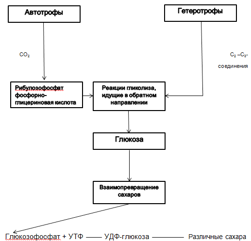
Author - L.B. Borisov, p. 51 "Medical Microbiology"
Figure 2.2.3. Lipid biosynthesis
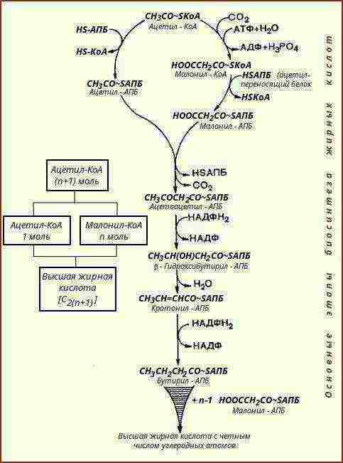
Table 2.2.4. Stages of energy metabolism - Catabolism.
|
№ |
Stages |
Characteristic |
Note |
|
Preparatory |
Molecules of disaccharides and polysaccharides, proteins break down into small molecules - glucose, glycerin and fatty acids, amino acids. Large nucleic acid molecules per nucleotide. |
At this stage, a small amount of energy is released, which is dissipated in the form of heat. |
|
|
Anoxic or incomplete or anaerobic or fermented or dissimilated. |
The substances formed at this stage with the participation of enzymes are further degraded. For example: glucose breaks down into two molecules of lactic acid and two molecules of ATP. |
ATP and H3PO4 are involved in glucose cleavage reactions. During the oxygen-free breakdown of glucose, 40% of the energy is stored in the ATP molecule in the form of a chemical bond, the rest is dissipated in the form of heat. In all cases of the breakdown of one glucose molecule, two ATP molecules are formed. |
|
|
The stage of aerobic respiration or oxygen breakdown. |
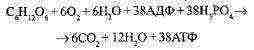
When oxygen is available to the cell, the substances formed during the previous stage are oxidized (broken down) to the final productsCO and HO. |
The total equation of aerobic respiration:
|
Scheme 2.2.4. Fermentation.
Fermentation metabolism -characterized by the formation of ATP by phosphorylation of substrates.
-
First (oxidation) = cleavage
-
Second (recovery)
Includes the conversion of glucose to pyruvic acid.
Includes hydrogen recovery for pyruvic acid recovery.
Pathways for the formation of pyruvic acid from carbohydrates
Scheme 2.2.5. Pyruvic acid.
Glycolytic pathway (Embden-Meyerhof-Parnassus pathway)
Entner-Dudorov path
Pentose phosphate pathway
Table 2.2.5. Fermentation.
|
№ |
Fermentation type |
Representatives |
Final product |
Notes (edit) |
|
Lactic acid |
|
Forms lactic acid from pyruvate |
In some cases (homoferment fermentation) only lactic acid is formed, in others also by-products. |
|
|
Formic acid |
|
Formic acid is one of the end products. (along with her - side) |
Some enterobacteriaceae break down formic acid to H2 and CO2 / |
|
|
Butyric acid |
|
Butyric acid and by-products |
Some types of clostridia, along with butyric and other acids, form butanol, acetone, etc. (then it is called acetone-butyl fermentation). |
|
|
Propionic acid |
|
Forms propionic acid from pyruvate |
Many bacteria ferment carbohydrates along with other foods to form ethyl alcohol. However, it is not a main product. |
Table 2.3.1. Protein synthesis system, ion exchange.
|
№ |
Item name |
Characteristic |
|
Ribosomal subunits 30S and 50S |
In the case of bacterial 70S ribosomes, the 50S subunit contains 23S rRNA (~ 3000 nucleotides in length) and the 30S subunit contains 16S rRNA (~ 1500 nucleotides in length); the large ribosomal subunit, in addition to the “long” rRNA, also contains one or two “short” rRNAs (5S rRNA of bacterial ribosomal subunits 50S or 5S and 5.8S rRNA of large ribosomal subunits of eukaryotes). (For more details, see Fig. 2.3.1.) |
|
|
Messenger RNA (mRNA) |
RNA containing information about the primary structure (amino acid sequence) of proteins |
|
|
A complete set of twenty aminoacyl-tRNAs, for the formation of which the corresponding amino acids are required, aminoacyl-tRNA synthetases, tRNA and ATP |
It is an amino acid, charged with energy and bound to tRNA, ready to be transported to the ribosome and incorporated into the polypeptide synthesized on it. |
|
|
Transport RNA (tRNA) |
Ribonucleic acid, the function of which is to transport amino acids to the site of protein synthesis. |
|
|
Protein initiation factors |
(in prokaryotes - IF-1, IF-2, IF-3) They got their name because they are involved in the organization of an active complex (708-complex) of 30S and 50S subunits, mRNA and initiator aminoacyl-tRNA (in prokaryotes, formylmethionyl -tRNA), which "starts" (initiates) the work of ribosomes - the translation of mRNA. |
|
|
Protein elongation factors |
(in prokaryotes - EF-Tu, EF-Ts, EF-G) Participate in the elongation (elongation) of the synthesized polypeptide chain (peptidil). Protein release factors (RF) provide codon-specific separation of the polypeptide from the ribosome and termination of protein synthesis. |
|
|
№ |
Item name |
Characteristic |
|
Protein termination factors |
(in prokaryotes - RF-1, RF-2, RF-3) |
|
|
Some other protein factors (associations, dissociation of subunits, release, etc.). |
Protein translation factors required for the functioning of the system |
|
|
Guanosine Triphosphate (GTP) |
For broadcasting, the participation of GTF is required. The need of the protein synthesizing system for GTP is very specific: it cannot be replaced by any of the other triphosphates. The cell spends more energy on protein biosynthesis than on the synthesis of any other biopolymer. The formation of each new peptide bond requires the cleavage of four high-energy bonds (ATP and GTP): two in order to load the tRNA molecule with an amino acid, and two more during elongation - one during the binding of aa-tRNA and the other during translocation. |
|
|
Inorganic cations at a certain concentration. |
To maintain the pH of the system within physiological limits. Ammonium ions are used by some bacteria to synthesize amino acids, potassium ions are used to bind tRNA to ribosomes. Iron and magnesium ions play the role of a cofactor in a number of enzymatic processes |
Figure 2.3.1. Schematic representation of the structures of prokaryotic and eukaryotic ribosomes.
Author - Korotyaev, p. 68 "Medical Microbiology"
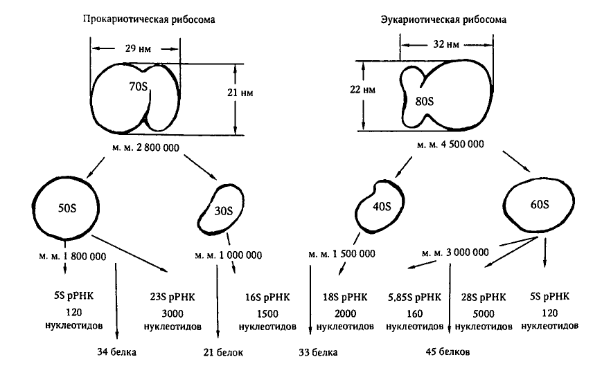
Table 2.3.2. Features of ion exchange in bacteria.
|
№ |
Peculiarity |
Characterized by: |
|
|
High osmotic pressure |
Due to the significant intracellular concentration of potassium ions in bacteria, a high osmotic pressure is maintained. |
||
|
Iron intake |
For a number of pathogenic and opportunistic bacteria (Escherichia, Shigella, etc.), iron consumption in the host's body is difficult due to its insolubility at neutral and slightly alkaline pH values. |
Siderophores -special substances that, by binding iron, make it soluble and transportable. |
|
|
Assimilation |
Bacteria actively assimilate SO2 / and PO34 + anions from the environment for the synthesis of compounds containing these elements (sulfur-containing amino acids, phospholipids, etc.). |
||
|
Jonah |
For the growth and reproduction of bacteria, mineral compounds are required - ions NH4 +, K +, Mg2 +, etc. (for more details, see Table 2.3.1.) |
||
Table 2.3.3. Ion exchange
|
№ |
Name of mineral compounds |
Function |
|
NH4 + (ammonium ions) |
Used by some bacteria to synthesize amino acids |
|
|
K + (potassium ions) |
|
|
|
Fe2 + (iron ions) |
|
|
|
Mg2 + (magnesium ions) |
||
|
SO42- (sulfate anion) |
Necessary for the synthesis of compounds containing these elements (sulfur-containing amino acids, phospholipids, etc.) |
|
|
PO43- (phosphate anion) |
Scheme 2.4.1. Energy metabolism.
For synthesis, bacteria need ...
-
Nutrients
-
Energy
Table 2.4.1. Energy metabolism (biological oxidation).
|
№ |
Process |
Necessary: |
|
Synthesis of structural components of microbial cells and maintenance of vital processes |
Adequate amount of energy. This need is met by biological oxidation, as a result of which ATP molecules are synthesized. |
|
|
Energy (ATP) |
Iron bacteria receive energy released during their direct oxidation of iron (Fe2 + to Fe3 +), which is used to fix CO2, bacteria metabolizing sulfur, provide themselves with energy due to the oxidation of sulfur-containing compounds. However, the vast majority of prokaryotes obtain energy through dehydrogenation. Energy is also received in the process of breathing (for a detailed table, see the corresponding section). |
Scheme 2.4. Biological oxidation in prokaryotes.
Decomposition of polymers into monomers
Stage I
Protein
Fats
Carbohydrates
glycerin and fatty acids
amino acids
monosaccharides
Decomposition under anoxic conditions
II stage
Formation of intermediate products
Oxidation under oxygen conditions to final products
Stage III
CO2
H2O
Table 2.4.2. Energy metabolism.
|
№ |
Concept |
Characteristic |
|
Essence of Energy Metabolism |
Providing the energy of cells necessary for the manifestation of life. |
|
|
ATF |
The ATP molecule is synthesized as a result of the transfer of an electron from its primary donor to the final acceptor. |
|
|
Breath |
|
|
|
Mobilizing energy |
Energy is mobilized in oxidation and reduction reactions. |
|
|
Oxidation reaction |
The ability of a substance to donate electrons (oxidize) |
|
|
Recovery Reaction |
The ability of a substance to attach electrons. |
|
|
Redox potential |
The ability of a substance to donate (oxidize) or receive (recover) electrons. (quantitative expression) |
Scheme 2.5. Synthesis.
SYNTHESIS
proteins
fat
carbohydrates
Table 2.5.1. Synthesis
|
№ |
Name |
Characteristic |
|
Cytoplasm |
The synthesis of the initial products occurs in the cytoplasm. |
|
|
Cytoplasmic membrane |
The initial products from the cytoplasm are transferred to the outer surface of the cytoplasmic membrane. |
|
|
Morphogenesis |
On the CPM, morphogenesis begins, that is, the formation of cell structures (capsules, cell walls, etc.) with the participation of enzymes. |
Table 2.5.1. Synthesis
|
№ |
Biosynthesis |
Of what |
Notes (edit) |
|
I |
Biosynthesis of carbohydrates |
Autotrophs synthesize glucose from CO2. Heterotrophs synthesize glucose from carbon-containing compounds. |
Calvin cycle (see diagram 2.2.1.) |
|
II |
Amino acid biosynthesis |
Most prokaryotes are able to synthesize all amino acids from:
|
The energy source is ATP. Pyruvate is formed in the glycolytic cycle. Auxotrophic microorganisms - consumed ready-made in the host's body. |
|
III |
Lipid biosynthesis |
Lipids are synthesized from simpler compounds - metabolic products of proteins and carbohydrates |
Acetyl-transfer proteins play an important role. Auxotrophic microorganisms - consume ready-made in the host's body or from nutrient media. |
Table 2.5.2. The main stages of protein biosynthesis.
|
№ |
Stages |
Characteristic |
Notes (edit) |
|
Transcription |
The process of RNA synthesis on genes. This is the process of rewriting information from DNA - gene to mRNA - gene. |
It is carried out using DNA - dependent RNA - polymerase. The transfer of information about the structure of the protein to ribosomes occurs with the help of mRNA. |
|
|
Broadcast (transmission) |
The process of own protein biosynthesis. The process of decoding the genetic code in mRNA and implementing it in the form of a polypeptide chain. |
Since each codon contains three nucleotides, the same genetic text can be read in three different ways (starting from the first, second and third nucleotides), that is, in three different reading frames. |
-
Note to the table: The primary structure of each protein is the sequence of amino acids in it.
Scheme 2.5.2. Electron transfer chains from the primary donor of hydrogen (electrons) to its final acceptor O2.
Organic matter
(primary electron donor)
NAD (- 0.32)
Flavoprotein (- 0.20)
Quinone (- 0, 07)
Cytochrome (+0.01)
Cytochrome C (+0.22)
Cytochrome A (+0.34)
O2 (+0.81)
final acceptor
Table 3.1. Classification of organisms by types of food.
|
№ |
Organogenic element |
Types of food |
Characteristic |
|
Carbon (C) |
|
They themselves synthesize all carbon-containing components of the cell from CO2. |
|
|
They cannot satisfy their needs with CO2, they use ready-made organic compounds. |
||
|
The food source is dead organic substrates. |
||
|
The food source is living tissues of animals and plants. |
||
|
Nitrogen (N) |
|
Satisfy their needs with atmospheric and mineral nitrogen |
|
|
Need ready-made organic nitrogenous compounds. |
||
|
Hydrogen (H) |
The main source is H2O |
||
|
Oxygen (O) |
|||
Table 3.1.2. Energy transformation
|
№ |
Classification |
Name |
Required: |
|
By energy source |
|
sunlight |
|
|
Redox reactions |
||
|
By electron donor |
|
Inorganic compounds (H2, H2S, NH3, Fe, etc.) |
|
|
Organic compounds |
Table 3.1.3. Carbon feeding methods
|
№ |
Energy source |
Electron donor |
Carbon feeding method |
|
Sunlight energy |
Inorganic compounds |
Photolithoheterotrophs |
|
|
Organic compounds |
Photoorganoheterotrophs |
||
|
Redox reactions |
Inorganic compounds |
Chemolithoheterotrophs |
|
|
Organic compounds |
Chemoorganoheterotrophs |
Table 3.2. Power mechanisms:
|
№ |
Mechanism |
Conditions |
Concentration gradient |
Energy costs |
Substrate specificity |
|
Passive diffusion |
The concentration of nutrients in the medium exceeds the concentration in the cell. |
By concentration gradient |
– |
– |
|
|
Facilitated diffusion |
Permease proteins are involved. |
By concentration gradient |
– |
+ |
|
|
Active transport |
Permease proteins are involved. |
Against the concentration gradient |
+ |
+ |
|
|
3A |
Translocation of chemical groups |
During the transfer process, chemical modification of nutrients occurs. |
Against the concentration gradient |
+ |
+ |
Table 3.3. Transport of nutrients from the bacterial cell.
|
№ |
Name |
Characteristic |
|
Phosphotransferase reaction |
Occurs when phosphorylation of the transferred molecule. |
|
|
Translational secretion |
In this case, the synthesized molecules must have a special leading amino acid sequence in order to attach to the membrane and form a channel through which protein molecules can escape into the environment. Thus, the toxins of tetanus, diphtheria and other molecules are released from the cells of the corresponding bacteria. |
|
|
Membrane budding |
The molecules formed in the cell are surrounded by a membrane vesicle, which is laced into the environment. |
Table 4. Growth.
|
№ |
Concept |
Definition of the concept. |
|
Growth |
An irreversible increase in the amount of living matter, most often due to cell division.If in multicellular organisms an increase in body size is usually observed, then in multicellular organisms the number of cells increases. But even in bacteria, an increase in the number of cells and an increase in cell mass should be distinguished. |
|
|
Factors affecting the growth of bacteria in vitro. |
Mycobacterium leprae is not capable of in vitro Chlamydia growth (including parasites)
|
|
|
Assessment of bacterial growth |
Growth quantification is usually carried out in liquid media where the growing bacteria form a homogeneous suspension. An increase in the number of cells is established by determining the concentration of bacteria in 1 ml, or the increase in cell mass is determined in weight units per unit volume. |
Growth factors
Lipids
Amino acids
Vitamins
Nitrogenous bases
Table 4.1. Growth factors
|
№ |
Growth factors |
Characteristic |
Function |
|
|
Amino acids |
|
Many microorganisms, especially bacteria, need one or more amino acids (one or more), since they cannot synthesize them on their own. Microorganisms of this kind are called auxotrophic for those amino acids or other compounds that they are unable to synthesize. |
||
|
Purine bases and their derivatives |
Nucleotides:
|
They are bacteria growth factors. Some types of mycoplasmas need nucleotides. Required for building nucleic acids. |
||
|
Pyrimidine bases and their derivatives |
Nucleotides
|
|||
|
№ |
Growth factors |
Characteristic |
Function |
|
|
Lipids |
|
Part of membrane lipids |
||
|
||||
|
Are components of phospholipids |
|||
|
In mycoplasmas, they are part of the cytoplasmic membrane |
|||
|
||||
|
Vitamins (mainly group B) |
|
Staphylococcus aureus, pneumococcus, Brucella |
||
|
All types of rod-shaped bacteria |
|||
|
Bifidobacteria and propionic acid |
|||
|
Some types of streptococci, tetanus bacilli |
|||
|
Yeast and nitrogen-fixing bacteria Rhizobium |
|||
|
Hemes - components of cytochromes |
Hemophilic bacteria, Mycobacterium tuberculosis |
|||
Table 5. Breathing.
|
№ |
Name |
Characteristic |
|
Breath |
Biological oxidation (enzymatic reactions) |
|
|
Base |
Breathing is based on redox reactions that lead to the formation of ATP, a universal accumulator of chemical energy. |
|
|
Processes |
When breathing, the following processes take place:
|
|
|
Aerobic breathing |
The final acceptor of hydrogen or electrons is molecular oxygen. |
|
|
Anaerobic breathing |
The acceptor of hydrogen or electrons is an inorganic compound - NO3-, SO42-, SO32-. |
|
|
Fermentation |
Organic compounds are acceptors of hydrogen or electrons. |
Table 5.1. Breathing classification.
|
№ |
Bacteria |
Characteristic |
Notes (edit) |
|
Strict anaerobes |
|
|
|
|
Strict aerobes |
|
Strict aerobes include, for example, representatives of the genus Pseudomonas |
|
|
№ |
Bacteria |
Characteristic |
Notes (edit) |
|
Facultative anaerobes |
|
Facultative anaerobes include enterobacteria and many yeasts that can switch from respiration in the presence of O2 to fermentation in the absence of O2. |
|
|
Microaerophiles |
A microorganism that requires, in contrast to strict anaerobes, for its growth the presence of oxygen in the atmosphere or nutrient medium, but in reduced concentrations compared to the oxygen content in ordinary air or in normal tissues of the host's body (in contrast to aerobes, for the growth of which normal oxygen content in the atmosphere or nutrient medium). Many microaerophiles are also capnophiles, that is, they require an increased concentration of carbon dioxide. |
In the laboratory, such organisms are easily cultivated in a "candle jar". A "candle jar" is a container into which a burning candle is introduced before being sealed with an airtight lid. The candle flame will burn until it is extinguished from a lack of oxygen, as a result of which an atmosphere saturated with carbon dioxide with a reduced oxygen content is formed in the can. |
Table 6. Characteristics of reproduction.
|
№ |
Name |
Characteristic |
|
Reproduction |
The term "propagation" refers to an increase in the number of cells in a population. Most prokaryotes reproduce by transverse division, some by budding. Fungi reproduce by sporulation. |
|
|
Where is going |
When a microbial cell multiplies, the most important processes occur in the nucleus (nucleoid), which contains all the genetic information in a double-stranded DNA molecule. |
Scheme 6. Dependence of the duration of generation on various factors.
Generation duration
Type of bacteria
Age
Population
Temperature
The composition of the nutrient medium
Table 6.1. Phases of bacterial reproduction.
|
№ |
Phase |
Characteristic |
|
I |
Initial stationary phase |
Lasts 1-2 hours. During this phase, the number of bacterial cells does not increase. |
|
II |
Lag phase (reproduction delay phase) |
It is characterized by the onset of intensive cell growth, but the rate of cell division remains low. |
|
III |
Log phase (logarithmic) |
Differs in the maximum rate of cell reproduction and an increase in the number of bacterial population exponentially |
|
IV |
Negative acceleration phase |
It is characterized by a lower activity of bacterial cells and a lengthening of the generation period. This occurs as a result of depletion of the nutrient medium, the accumulation of metabolic products in it and oxygen deficiency. |
|
V |
Stationary phase |
It is characterized by a balance between the number of dead, newly formed and dormant cells. |
|
VI |
Doom phase |
It occurs at a constant rate and is replaced by UP-USH phases of decreasing the rate of cell death. |
Scheme 7. Requirements for culture media.
Requirements
Viscosity
Humidity
Sterility
Nutritional value
Transparency
Isotonicity
pH of the environment
Table 7. Reproduction of bacteria on nutrient media.
|
№ |
Nutrient medium |
Characteristic |
|
|
Dense nutrient media |
On dense nutrient media, bacteria form colonies - clusters of cells. |
||
|
S - a type (smooth - smooth and shiny) Round, with an even edge, smooth, convex. |
R - a type (rough - rough, uneven) Irregular in shape with jagged edges, rough, dented. |
||
|
Liquid culture media |
|
||
Table 7.1. Classification of culture media.
|
№ |
Classification |
Views |
Examples of |
|
By composition |
Simple |
|
|
|
Complex |
|
||
|
By appointment |
The main |
|
|
|
Elective |
|
||
|
Differential - diagnostic |
|
||
|
Special |
|
||
|
By consistency |
Dense |
|
|
|
Liquid |
|
||
|
Semi-liquid |
|
||
|
By origin |
Natural |
|
|
|
Semi-synthetic |
|
||
|
Synthetic |
|
Table 7.2. Principles of isolation of pure cell culture.
|
Mechanical principle |
Biological principle |
|
METHODS 1. Fractional dilutions of L. Pasteur 2. Plate dilutions R. Koch 3. Surface crops Drigalsky 4. Surface strokes |
METHODS Consider: a - type of breathing (Fortner's method); b - mobility (Shukevich's method); c - acid resistance; d - sporulation; d - temperature optimum; e - selective sensitivity of laboratory animals to bacteria |
Table 7.2.1. Stages of isolation of pure cell culture.
|
№ |
Stage |
Characteristic |
|
Stage 1 research |
Take away pathological material. It is studied - appearance, consistency, color, smell and other signs, a smear is prepared, painted and examined under a microscope. |
|
|
Stage 2 research |
On the surface of a dense nutrient medium, microorganisms form a continuous, dense growth or isolated colonies.The colony - These are the accumulations of bacteria visible to the naked eye on the surface or in the thickness of the nutrient medium. As a rule, each colony is formed from the descendants of one microbial cell (clones), therefore their composition is quite homogeneous. Features of the growth of bacteria on nutrient media are a manifestation of their cultural properties. |
|
|
Stage 3 research |
The nature of the growth of a pure culture of microorganisms is studied and its identification is carried out. |
Table 7.3. Identification of bacteria.
|
№ |
Name |
Characteristic |
|
Biochemical identification |
Determination of the type of pathogen by its biochemical properties |
|
|
Serological identification |
In order to establish the species of bacteria, their antigenic structure is often studied, that is, they are identified by antigenic properties. |
|
|
Identification by biological properties |
Sometimes bacteria are identified by infecting laboratory animals with a pure culture and observing the changes that pathogens cause in the body. |
|
|
Cultural identification |
Determination of the type of pathogens by their cultural characteristics |
|
|
Morphological identification |
Determination of the type of bacteria by their morphological characteristics |
Rating control tests
-
Which of the processes is not related to the physiology of bacteria?
-
Growth
-
Reproduction
-
Mutation
-
Food
-
What substances make up 40 - 80% of the dry mass of a bacterial cell?
-
Carbohydrates
-
Protein
-
Fats
-
Nucleic acids
-
What classes of enzymes are synthesized by microorganisms?
-
Oxy reductase
-
All classes
-
Transferases
-
Ligases
-
Enzymes, the concentration of which in the cell sharply increases in response to the appearance of an inducer substrate in the medium?
-
Iiducible
-
Constitutional
-
Repressive
-
Multienzyme complexes
-
An enzyme of pathogenicity secreted by Staphylococcus aureus?
-
Neuraminidase
-
Hyaluronidase
-
Lecithinase
-
Fibrinolysin
-
Do proteolytic enzymes perform a function?
-
Protein breakdown
-
Breaking down fats
-
Breakdown of carbohydrates
-
Alkali formation
-
Fermentation of Enterobacteriaceae?
-
Lactic acid
-
Formic acid
-
Propionic acid
-
Butyric acid
-
What mineral compounds are used to bind t-RNA to ribosomes?
-
NH4
-
K +
-
Fe2 +
-
Mg2 +
-
Biological oxidation is ...?
-
Food
-
Reproduction
-
Breath
-
Cell death
-
What substances themselves synthesize all carbon-containing components of the cell from CO2.
-
Prototrophs
-
Heterotrophs
-
Autotrophs
-
Saprophytes
-
Culture media differ:
-
By composition
-
By consistency
-
By appointment
-
All of the above
-
The reproductive phase, which is characterized by a balance between the number of dead, newly formed and dormant cells?
-
Lag phase
-
Log phase
-
Negative acceleration phase
-
Stationary phase
-
Generation duration depends on?
-
Species
-
Age
-
Populations
-
All of the above
-
In order to establish the species of bacteria, their antigenic structure is often studied, that is, identification is carried out, which one?
-
Biological
-
Morphological
-
Serological
-
Biochemical
-
Drygalski's surface sowing method is referred to as ...?
-
Mechanical principles of isolation of pure culture
-
Biological principles of isolation of pure culture
Bibliography
1. Borisov LB Medical microbiology, virology, immunology: a textbook for honey. universities. - M .: LLC "Medical Information Agency", 2005.
2. Pozdeev OK Medical microbiology: a textbook for honey. universities. - M .: GEOTAR-MED, 2005.
3. Korotyaev AI, Babichev SA Medical microbiology, immunology and virology / textbook for honey. universities. - SPb .: SpetsLit, 2000.
4. Vorobiev A.A., Bykov A.S., Pashkov E.P., Rybakova A.M. Microbiology: textbook. - M .: Medicine, 2003.
5. Medical microbiology, virology and immunology: textbook / ed. V.V. Zvereva, M.N.Boychenko. - M .: GEOTar-Media, 2014.
6. Guide to practical training in medical microbiology, virology and immunology / ed. V.V. Teza. - M .: Medicine, 2002.
Content
Introduction 6
The composition of bacteria from the point of view of their physiology. 7
Metabolism 14
Nutrition (transport of nutrients) 25
Height 29
Breath 31
Breeding 34
Microbial communities 37
APPENDICES 49
Tests 102
References 105
…………..
…………
Some human diseases are associated with exposure to microbes on the body. When such diseases occur, complex changes occur in the human body, protective functions are mobilized, aimed at combating the trapped microbes.
Among the huge number of microorganisms, there are those that are capable of causing diseases in humans, animals and plants. They are called pathogenic or disease-causing. Specificity is characteristic of pathogenic microorganisms - each type of them is capable of causing only a certain disease with all its characteristic signs. Most pathogenic microorganisms are microbes and parasites, since they are able to live off the substances of a living organism.
Pathogenic microbes produce special substances - toxins that poison the body and cause a painful condition. The ability of microorganisms to cause disease is called pathogenicity. It can manifest itself to varying degrees. The degree of pathogenicity is called virulence.The virulence of microbes can increase or decrease both in natural and experimental conditions.
Under the term "infection»Understand the process of interaction of microbes with the human body, as a result of which an infectious disease occurs. Sources of infection are, first of all, sick people and animals that release pathogens into the external environment, as well as people and animals who have recovered from illness, in whose body pathogenic microbes continue to remain for some time (sometimes very long) after recovery. People and animals that release pathogenic microbes after recovery are called bacteria carriers or bacteria excretors. People who are not sick can also be bacteria carriers. The pathogenic microbes isolated by the sick organism enter the air, soil, water, surrounding objects and food, where they can remain viable for more or less a long time, depending on the type of microbes.
Pathogenic microbes enter the human body in various ways: through direct contact with a sick person, through the air with the smallest droplets of mucus and saliva secreted by the patient when coughing or sneezing (droplet infection). Microbes - the causative agents of some diseases - are excreted by patients with feces and urine. Such microbes enter the body of a healthy person through dirty hands that have not been washed after using the toilet. Contaminated hands also spread pathogens onto food. The causative agents of intestinal infections also penetrate into the healthy body when contaminated water is consumed.
Insects and rodents are often carriers of infectious diseases. Flies can carry the causative agents of typhoid fever and dysentery, lice - typhus, some types of mosquitoes - malaria, etc. Mice and rats are distributors of causative agents of plague, tularemia, anthrax.
The penetration of pathogenic microbes into the human body does not always lead to its disease. In the onset and course of the disease, the properties of pathogenic microbes that have penetrated the body, their number and activity, as well as the place of their introduction into the body and the state of the body itself play an important role.
For the reproduction of pathogenic microorganisms, favorable conditions are needed, and they appear when the human body is weakened and its protective ability is reduced (due to hypothermia, starvation, etc.). Susceptibility to infections also depends on age (in children and the elderly, it is higher than in adults).
From the moment pathogenic microbes enter the human body until the signs of the disease appear, a certain period of time usually passes - it is called the latent period of the disease, or incubation. During this time, there is a multiplication of pathogenic microbes in the body and the accumulation of harmful products of their vital activity. The duration of the incubation period for different diseases is not the same. It ranges from several days (flu, tetanus, dysentery) to several weeks (typhus, typhoid fever), and sometimes reaches several months (rabies) and even years (leprosy).
If you find an error, please select a piece of text and press Ctrl + Enter.
Culture media - substrates consisting of components that provide the necessary conditions for the cultivation of microorganisms or the accumulation of their waste products
.
Culture media differ in purpose, consistency and composition. According to their purpose, they are conventionally divided into two main groups - diagnostic and production environments. Diagnostic culture media include five subgroups: media for growing a wide range of microorganisms; medium for the isolation of a specific pathogen; differential media allowing to distinguish between certain types of microorganisms; media for the identification of microorganisms and storage media intended for the enrichment of a specific type of microorganisms. Production includes P. with. used in the industrial production of biological medicinal products (bacterial vaccines, toxoids, etc.) and quality control.Culture media for the production of bacterial preparations, in contrast to diagnostic media, should not contain impurities harmful to humans and have a negative effect on the production process (without interfering, in particular, with the removal of products of microbial metabolism, ballast organic and mineral substances from the prepared preparations).
By consistency, liquid, dense and semi-liquid media are distinguished. Solid and semi-liquid nutrient media are prepared from liquid media by adding agar or (less often) gelatin to them. Agar agar is a polysaccharide obtained from certain varieties of seaweed. Agar is usually introduced into P. with. in a concentration of 1-2% (in the manufacture of semi-liquid media - 0.2-0.4%), gelatin - 10-15%. At t ° 25-30 ° gelatinous media melt, so microorganisms are grown on them mainly at room temperature. Clotted blood serum, rolled eggs, potatoes and media containing 1.5% silica gel are also used as solid media.
In terms of composition, culture media are divided into simple and complex. Simple P. with. provide the nutritional needs of most pathogenic microorganisms (mesopatamia broth, mesopatamia agar, Hottinger's broth and agar, nutritious gelatin, peptone water, etc.). Complex media include special media for microbes that do not grow on simple nutrient media. Such media are prepared by adding blood, serum, carbohydrates and other substances necessary for the reproduction of a particular microorganism to simple media. Complex P. with. are also differential diagnostic media, for example, Giss media with carbohydrates and an indicator, used to determine the species of the microbe under study by its enzymatic activity. Some complex media, the so-called selective, or elective, are used for the targeted isolation of one or several types of microorganisms by creating optimal conditions for their growth and inhibition of the growth of the accompanying microflora. For example, bismuth sulfite agar is a strictly selective medium for the isolation of Salmonella, agar and Levin's medium are weakly selective media for the isolation of enterobacteria.
Depending on the composition of the initial components, synthetic, semi-synthetic and natural media are also distinguished. Synthetic media, the components of which are precisely known, and to a somewhat lesser extent semi-synthetic, are convenient and are used mainly for studying the physiological processes of microorganisms. The use of media of this type makes it possible to determine the minimum requirements of individual microorganisms for nutrients and, on this basis, create nutrient media containing only those compounds that are necessary for the growth of a given microbe. The advantage of synthetic culture media is their standardization, however, the use of these media is limited due to the high cost and complexity of the composition of many of them (they often contain up to 40 or more ingredients). In addition, they are more sensitive to imbalances between some of the P.'s components. , especially amino acids, and the multiplication of microorganisms on such media is more easily suppressed by excessive aeration or toxic cations.
In microbiological practice, media of natural origin continue to be widely used, the chemical composition of which is not well known. The basis of such media is made from various raw materials of animal or vegetable origin: meat and its substitutes, fish, animal blood, casein, yeast, potatoes, soybeans, etc. much of the environments themselves.
Along with protein bases, nutrient media should contain ash elements (phosphorus, sulfur, calcium, magnesium, iron) and trace elements (boron, molybdenum, cobalt, manganese, zinc, nickel, copper, chlorine, sodium, silicon, etc.). These substances are necessary for many biosynthetic processes in bacteria, but the need for them in different types of microorganisms is not the same. For example, manganese and iron are required for E. coli and plague pathogens, and potassium for lactic acid bacteria. Manganese, calcium, magnesium, potassium and iron stimulate the growth of the anthrax bacillus, and magnesium, iron and manganese stimulate the growth of brucella.
For the cultivation of many pathogenic microorganisms, the presence in the medium of the so-called growth factors, represented primarily by water-soluble vitamins, is necessary. Although bacteria do not use these substances as a plastic or energetic material, nevertheless they are indispensable components of nutrient media, because their absence inhibits the formation of many enzymes. Certain organic acids, purine and pyrimidine bases and amino acids can also be growth factors. Certain amino acids (L-cystine, D-pyroglutamic acid, etc.) are able to stimulate the growth of some microorganisms and, conversely, inhibit the growth of others.
Culture media must contain all the chemical elements necessary for the grown microorganisms in an easily assimilable form and in optimal quantities. The source of carbon in P. s. usually there are individual carbohydrates, salts of organic acids, as well as carbon, which is part of nitrogen-containing compounds (proteins, peptones, amino acids, etc.). Nitrogen requirements are met if there is a native animal protein in the media (blood, serum, ascitic fluid, etc.), peptones, amino acids, ammonium salts and other nitrogen-containing substances (purine and pyrimidine bases, urea, etc.). IN nutrient media make mineral salts necessary for microorganisms. Microelements required by bacteria in negligible amounts usually get into nutrient media either with water, which is used to prepare the medium, or with the raw materials that are part of the nutrient medium. Growth factors enter the environment with various dialysates, extracts and autolysates in the form of amino acids, peptides, purine and pyrimidine bases and vitamins.
Protein-containing raw materials used for the preparation of culture media are hydrolyzed using various enzymes (pepsin, trypsin, pancreatin, papain, fungal proteases, etc.) or acids (less often alkalis). The purpose of hydrolysis is to dissolve and break down the protein with the formation of nitrogenous compounds assimilated by the microbial cell: peptones, polypeptides, amino acids, which are part of the hydrolysates obtained in this way. To meet the nutritional needs of each specific type of microorganism, these or those hydrolysates are used, depending on their chemical composition, or in the composition of P. with. several hydrolysates are introduced at the same time in the used ratios.
Designed P. with. in its composition and physicochemical properties, it must correspond to the conditions of the natural habitation of microorganisms. The differences in their nutritional needs are so diverse that they exclude the possibility of creating a universal P. with. Therefore, modern principles of media design are based on a comprehensive study of the nutritional requirements of microorganisms.
It is necessary that nutrient media not only contain nutrients necessary for microbes, but also have an optimal concentration of hydrogen and hydroxyl ions. Most pathogenic microorganisms grow better on nutrient media with a slightly alkaline reaction (pH 7.2-7.4).The exceptions are Vibrio cholerae, the optimum growth of which is in the alkaline zone (pH 8.5-9.0), and the causative agent of tuberculosis, which requires a slightly acidic reaction (pH 6.2-6.8). To prevent a change in pH during the cultivation of microorganisms in P. with. add phosphate buffers - a mixture of mono- and disubstituted potassium phosphates, the concentration of which in the medium should not exceed 0.5%.
Culture media must have sufficient humidity and be isotonic for the microbial cell, which ensures the normal course of the most important physical and chemical processes in it. For most microorganisms, the optimal environment is 0.5% sodium chloride solution. One of the essential requirements for culture media is their sterility, which makes it possible to grow pure cultures of microbes.
An important role in P.'s receipt by page. proper quality belongs to the methods of their control. For nutrient media used in production, the main quality indicator is the media efficiency (yield), i.e. the ability to accumulate the maximum amount of biomass of microorganisms of full properties or products of their biosynthesis (toxins, etc.). In assessing diagnostic nutrient media, the leading criterion is the sensitivity indicator - the ability to ensure the growth of microorganisms in the maximum dilutions of the pathogen culture. Depending on the purpose of the nutrient media, when assessing their quality, other indicators are also used - the stability of the main properties of the grown microorganisms and the rate of their growth, the severity of the differentiating properties, etc.
When solving the quality issues of nutrient media, important importance is attached to their standardization, which is facilitated by the production of media in the form of dry preparations. In the USSR, industrial production of dry media for various purposes has been established: simple, selective, differential diagnostic and special.
Bibliography: Yu.A. Kozlov Culture media in medical microbiology, M., 1950, bibliogr .; Laboratory research methods in the clinic, ed. V.V. Menshikov, s. 315, 343, M., 1987; Meinell J. and Meinell E. Experimental Microbiology, trans. from English, p. 46, M., 1967; Microbiological research methods in infectious diseases, ed. G.Ya. Sinai and O.G. Birger, s. 64, M., 1949; Enterobacteriaceae, ed. IN AND. Pokrovsky, p. 258, M., 1985.
Attention! Article 'Culture media'Is provided for informational purposes only and should not be used for self-medication