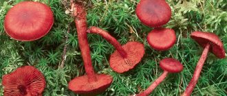
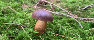
Areas of abnormal spider webs growth.
Anomalous cobwebs settle in small groups, can occur singly. Mostly they grow in deciduous forests and conifers. They grow on the ground, or on a litter of leaves and needles. Fruiting is observed from August to September.

Anomalous webcaps are common in Bulgaria, Germany, Belgium, Austria, Norway, Switzerland, Great Britain, Sweden, France, Belarus and Estonia. Also, this species is found in the United States, Morocco and the Greenland Islands. In addition, anomalous spider webs are known in some regions of Russia, for example, in the Chelyabinsk, Irkutsk, Amur, Tver and Yaroslavl regions, in the Primorsky, Krasnoyarsk and Khabarovsk territories, as well as in Karelia.

Evaluation of the edibility of the spiderweb abnormal.
The nutritional properties of this genus are poorly understood, but scientists believe that the abnormal cobweb belongs to an inedible species that cannot be collected and eaten.
Related species.
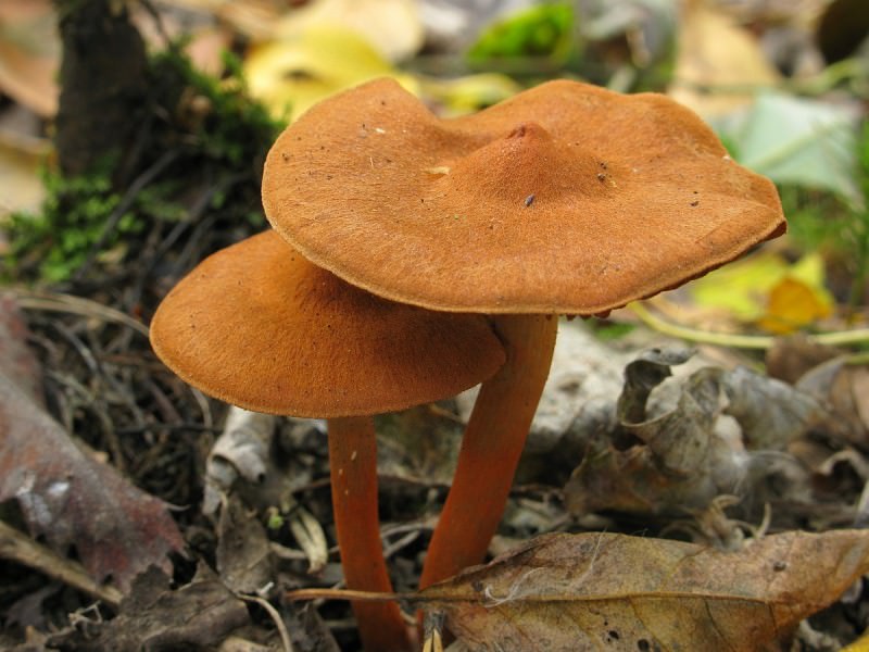
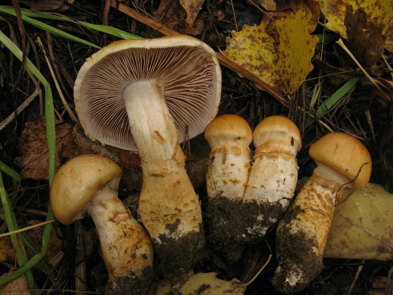
The cinnamon webcap is also an inedible mushroom. His cap varies from hemispherical to open. It is dry to the touch. The structure of the cap is fibrous. The color of the cap is yellow-brown or yellow-brown. The leg is cylindrical, at first there is pulp in it, but gradually it becomes hollow. The coloring of the leg is yellow-brown. The pulp is without a noticeable smell and taste, yellow in color.

Fruiting of cinnamon spiderwebs is observed from August to September. This species grows in mixed forests and conifers. Occurs singly or in groups.
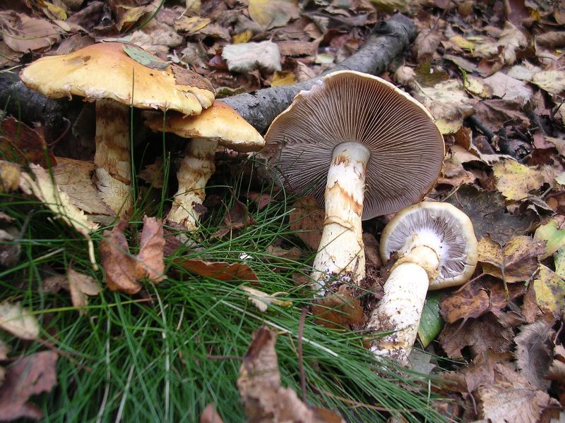
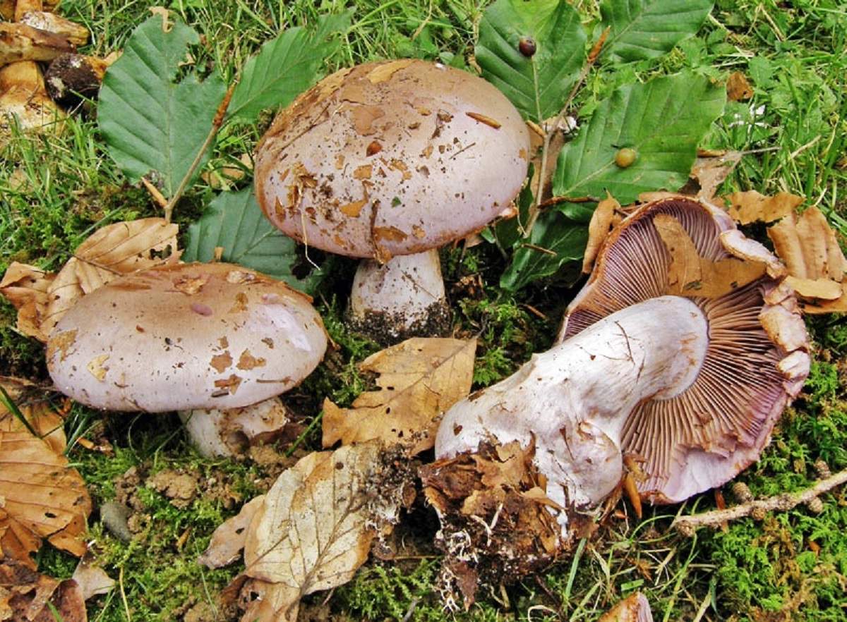
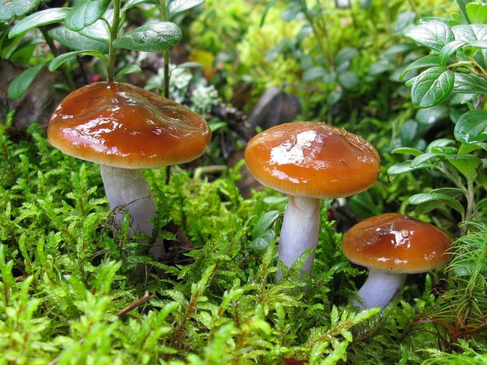
The cobweb is also an inedible relative of the abnormal cobweb. The cap is hemispherical at first, later becomes convex. At high humidity, it becomes covered with mucus. The color of the fruit bodies is at first light purple, and then turns red-brown. The flesh tastes fresh, and its smell is mildly unpleasant. The flesh is white in color, sometimes with a purple tint. The leg is widened in the lower part, light purple or brownish in color; the remains of the bedspread can be seen on its surface.

This is a widespread type of spider webs in the Eurasian zone. Most often, fruiting bodies are found in large groups. They settle in mixed or deciduous forests. They find them under beeches and oaks.
Definitioner
- rare (rare smell)
-
In mycology, a rare smell, English. "Raphanoid", is interpreted very loosely and often denotes any smell of raw root vegetables, including potato, ie. not necessarily as sharp, sharp, and crisp as black or white radish.
- Basidia (Basidia)
-
Lat. Basidia. A specialized structure of sexual reproduction in fungi, inherent only in Basidiomycetes. Basidia are terminal (end) elements of hyphae of various shapes and sizes, on which spores develop exogenously (outside).
Basidia are diverse in structure and method of attachment to hyphae.
According to the position relative to the axis of the hypha, to which they are attached, three types of basidia are distinguished:
Apical basidia are formed from the terminal cell of the hypha and are located parallel to its axis.
Pleurobasidia are formed from lateral processes and are located perpendicular to the axis of the hypha, which continues to grow and can form new processes with basidia.
Subasidia are formed from a lateral process, turned perpendicular to the axis of the hypha, which, after the formation of one basidium, stops its growth.
Based on morphology:
Holobasidia - unicellular basidia, not divided by septa (see Fig. A, D.).
Phragmobasidia are divided by transverse or vertical septa, usually into four cells (see Fig. B, C).
By type of development:
Heterobasidia consists of two parts - hypobasidia and epibasidia developing from it, with or without partitions (see Fig. C, B) (see Fig. D).
Homobasidia is not divided into hypo- and epibasidia and in all cases is considered holobasidia (Fig. A).
Basidia is the place of karyogamy, meiosis and the formation of basidiospores. Homobasidia, as a rule, is not functionally divided, and meiosis follows karyogamy in it. However, basidia can be divided into probasidia - the site of karyogamy and metabasidia - the site of meiosis. Probasidium is often a dormant spore, for example in rust fungi. In such cases, probazidia grows with metabasidia, in which meiosis occurs and on which basidiospores are formed (see Fig. E).
See Karyogamy, Meiosis, Gifa.
- Pileipellis
-
Lat. Pileipellis, skin - differentiated surface layer of the cap of agaricoid basidiomycetes. The structure of the skin in most cases differs from the inner flesh of the cap and may have a different structure. The structural features of pileipellis are often used as diagnostic features in descriptions of fungi species.
According to their structure, they are divided into four main types: cutis, trichoderma, hymeniderma and epithelium.
See Agaricoid fungi, Basidiomycete, Cutis, Trichoderma, Gimeniderm, Epithelium.
Description of the spider web is abnormal.
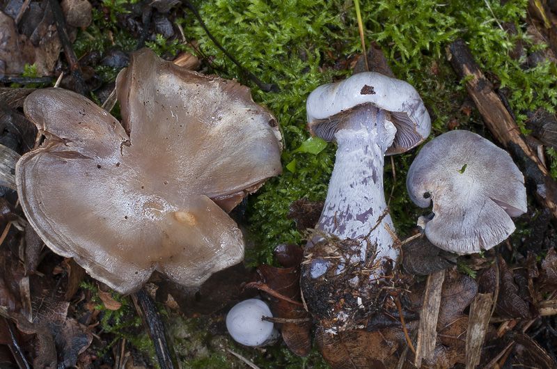
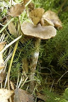
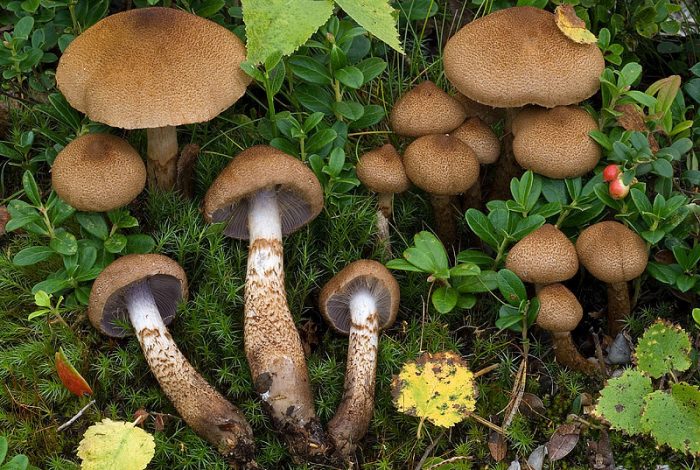
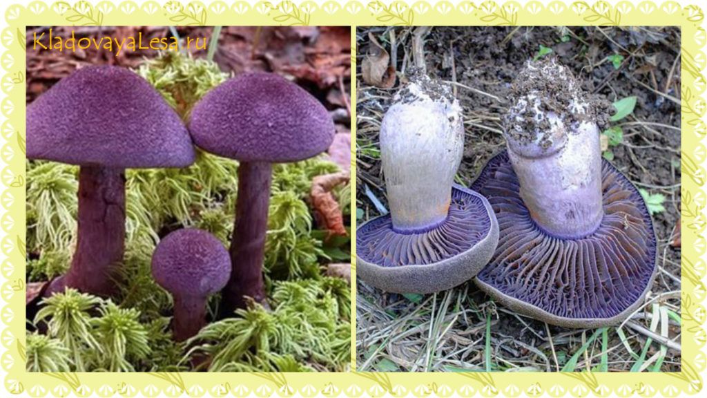
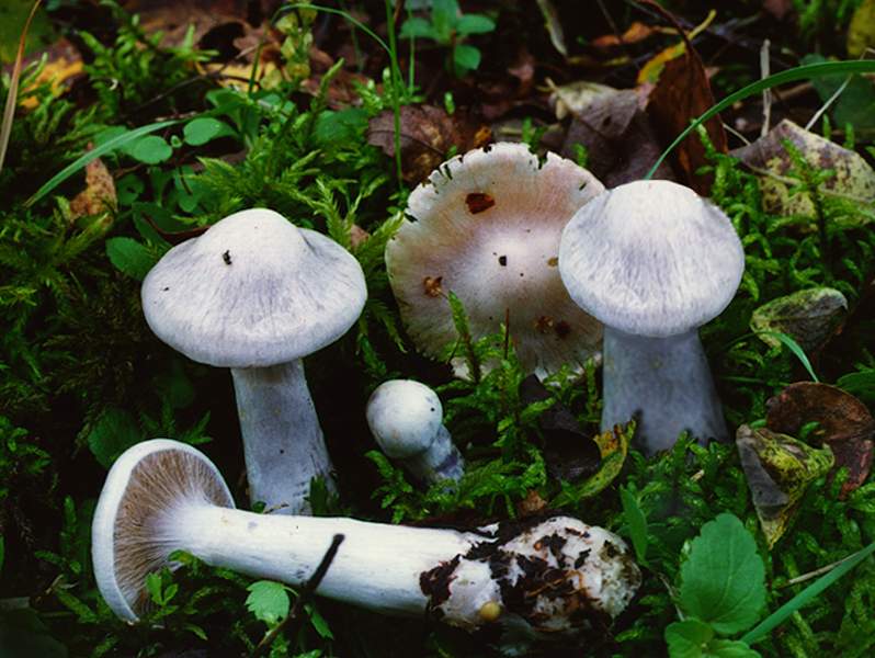
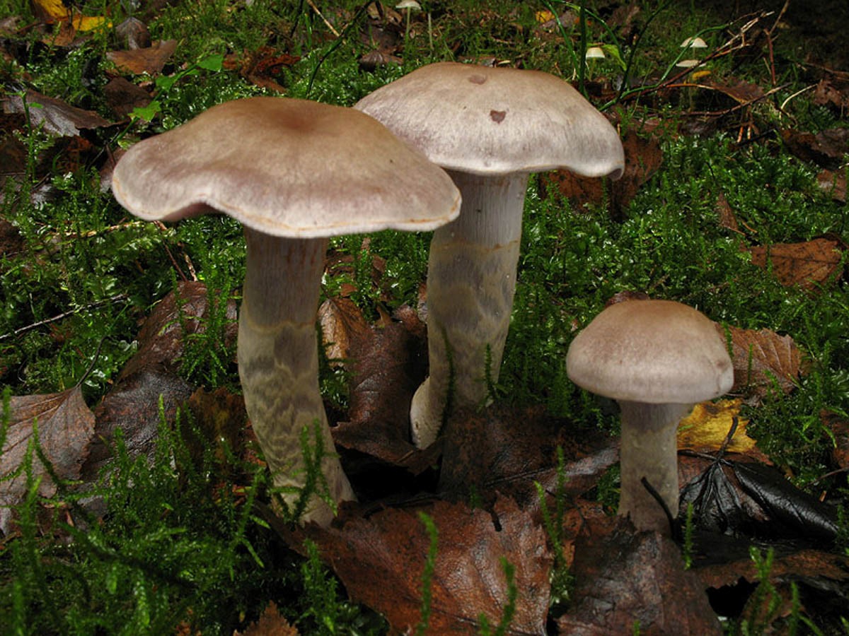
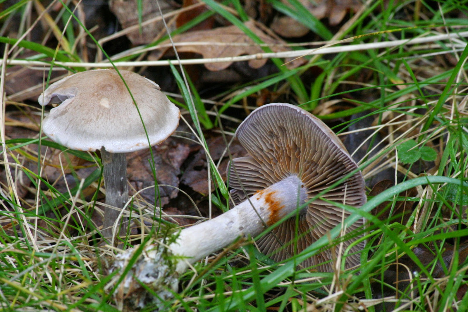
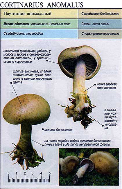
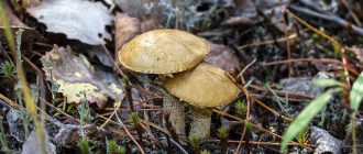
The fruit body of the abnormal spider web consists of a leg and a cap. Initially, his hat is convex, but in adulthood it becomes flat. The hat is smooth, silky and dry to the touch. The color of the cap at a young age is gray or grayish-brown, and its edges are bluish-purple, gradually its color becomes brown or red-brown.

The mushroom leg is cylindrical in shape, at the base it has a thickening. Its length is 7-10 centimeters, and the girth reaches 1 centimeter. In young spider webs, the legs are full, while in mature ones they become empty from the inside. The color of the leg is whitish, while a lilac or brown tint is observed. On the surface of the leg, fibrous remnants of a frequent bedspread are noticeable; they are lighter in color than the main background.
The mushroom pulp of the abnormal webcap is well developed. The color of the pulp is whitish, and in the leg it has a purple tint. The flesh tastes soft and has no smell.

The hymenophore of the spider web is anomalous lamellar. The plates are wide, they are often located, grow to the inner surface of the cap. In young mushrooms, the color of the plates is gray-lilac, but when the fruiting bodies ripen, the plates become brown-rusty. The records contain spores. The shape of the spores is broadly oval; they are pointed at the ends. The color of the spores is light yellow. Their surface is warty.

