Season and habitat of the mushroom
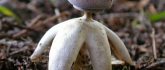
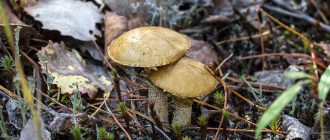
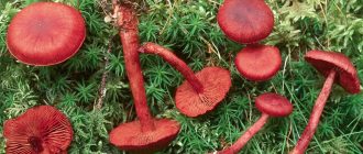
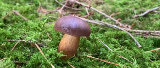
Previously, the Pisolíthus tinctorius mushroom was classified as a cosmopolitan, and it could be found almost everywhere, except in the regions located beyond the Arctic Circle. Now the boundaries of the habitat of this fungus are being revised, since some of its subspecies, growing, for example, in the southern hemisphere and the tropics, are classified as separate species. Based on this information, it can be said that pisolithus is found in the Holarctic, but its varieties found in South Africa and Asia, Central Africa, Australia, New Zealand, most likely belong to related types. On the territory of Russia, pisolithus can be seen in Western Siberia, in the Far East and in the Caucasus. The period of the most active fruiting occurs in summer and early autumn. Grows either singly or in small groups.
The dye pisolithus grows mainly on acidic and poor soils, in forest clearings, gradually overgrowing, on green dumps and gradually overgrown quarries. However, these fungi can never be seen on limestone-type soils. It is extremely rare that it grows in forests that are practically untouched by humans. Can form mycorrhiza with birch and coniferous trees. It is a mycorrhizal agent with eucalyptus, poplars and oaks.
Cellular slime molds are very different.
As you can see, the strategies of aggregation and plasmodation have little in common. Therefore, biologists have long assumed that cellular and plasmodial slime molds are unrelated to each other. In the 1970s, one more circumstance became clear: the strategy of cellular slime molds arose repeatedly in the course of evolution, in a variety of organisms (L. S. Olive, 1970. The Mycetozoa: a revised classification). Today we know that this extravagant path of development is practiced by at least six unrelated groups. Among them dictyostelids and copromixides belong to the supergroup Amoebozoa and are related to the common amoeba from the school textbook, phoniculids belong to Opisthokonta and are considered close relatives of real mushrooms, and arazids belong to Discoba and are distantly related to euglena and trypanosomes (see Accidentally discovered flagellate updates the eukaryotic system, "Elements", 02/06/2018). The fifth group, which has mastered aggregation, is, oddly enough, ciliates (supergroup Alveolata), although among them fruiting bodies form only one species, Sorogena stoianovitchiae (see S. Sharpe et al., 2015. Timing the origins of multicellular eukaryotes through phylogenomics and relaxed molecular clock analyzes). Finally, aggregation with the formation of fruiting bodies is widespread inmyxobacteria - prokaryotes from the Proteobacteria division. Mobile "pseudoplasmodia" of these organisms, the so-called swarms, are able to lyse and eat other bacteria. However, myxobacteria are not capable of phagocytosis, which excludes them from the list of "classic" slime molds.
An important conclusion follows from the above: the developmental path characteristic of cellular slime molds is not an exotic evolutionary accident, but a completely common and successful strategy that has been repeatedly used by a variety of organisms. It is pertinent to note here that the mycelial structure was independently "invented" by only two taxonomic groups (real and false fungi), and the multicellularity of the animal type, apparently, generally appeared on Earth only once (see, for example: S. Yastrebov, 2016. Cambrian explosion). Thus, the appearance of cellular slime molds is a more natural event than the appearance of animals.
Crowded pseudombrophila (Pseudombrophila aggregata)
Synonyms:
- Nanfeldia crowded
- Nannfeldtiella aggregate

The crowded pseudombrophilia is a species with a rather confusing history.
Described as Nannfeldtiella aggregata Eckbl. (Finn-Egil Ekblad (Norv.Finn-Egil Eckblad, 1923-2000) - Norwegian mycologist, specialist in discomycetes) in 1968 as a monotypic species Nannfeldtiella (Nannfeldtia) in the Sarcoscyphaceae family (Sarcoscyphae). Further research indicated that the species should be placed in the Pyronemataceae.
Please note that in almost all photographs used for illustration, there are two types of mushrooms. Bright orange small "buttons" are Byssonectria terrestris
Larger brown "cups" are just the crowded Pseudombrophila. The fact is that these two species always grow together, apparently forming a symbiosis.
Description
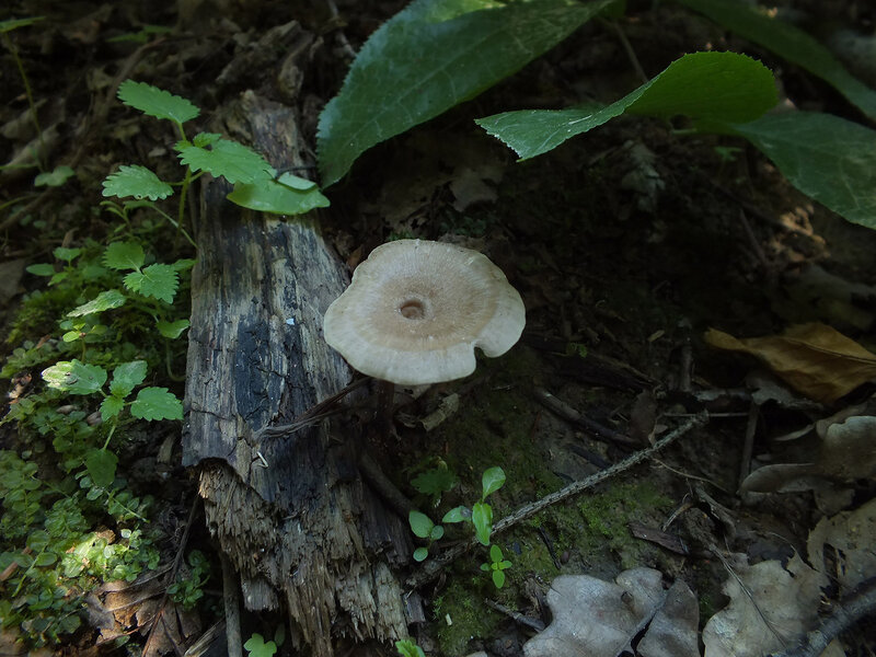
Fruit body: at first spherical, from 0.5 to 1 centimeter in diameter, with a pubescent surface, then stretches slightly, opens up, acquiring a cupped shape, light brownish, the color of coffee with milk or brownish with a lilac tint, with a well-pronounced darker ribbed edge. With age, it expands to the saucer-shaped, while maintaining the "ribbing" of the edge.

In adult fruiting bodies, the size can be up to one and a half centimeters in diameter. The color is light chestnut, brownish, brown, lilac or purple shades may be present. The inner side is darker, smoother and more shiny. The outer side is lighter, retains the edge. The integumentary hairs are sparse from above, rather thick downward, oddly curved, 0.3-0.7 microns thick.

Stem: absent or very short, weakly expressed.
Flesh: the mushroom is rather "fleshy" (in proportion to its size), the flesh is dense, without much taste or smell.
Microscopy
Asci are 8-spore, all eight spores mature.
Spores 14.0-18.0 x 6.5-8.0 μm, fusiform, ornamented.
Ecology
In forests of various types, on leaf litter and on small rotting twigs, in the vicinity of ground Bissonektria. It is considered an "ammonia" fungus, as it grows in places where moose urine is present in the ground.
Edibility
Given the small size of the fruiting bodies and taking into account the specifics of growth (on the urine of moose), there are probably not so many who want to experiment with edibility.
No data on toxicity
Similar species
Several species of Pseudombrophila are indicated, growing together with some kind of Byssonectria sp. They differ at the microscopic level, the size of spores and their number in asci and the thickness of the integumentary hairs, at the ecology level - the place of growth, namely, on the excrement of which herbivore animal they have grown. Unfortunately, the usual mushroom picker or photographer has little or no way to distinguish between these species.
Photo: Alexander, Andrey, Sergey.
Literature used: Harmaja, H. 1979. Studies on vernal species of Gyromitra and Pseudombrophila (syn. Nannfeldtiella). Acta Botanica Fennica. 16 (3): 159-162
Ceratiomyxa dwarf shrub (Ceratiomyxa fruticulosa)
Synonyms:
- Keratiomix subshrub
- Keratomix subshrub
- Byssus fruticulosa

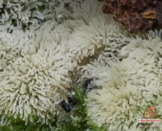
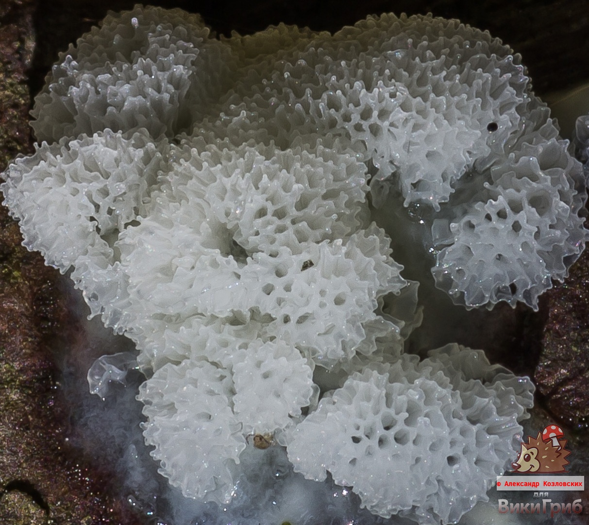
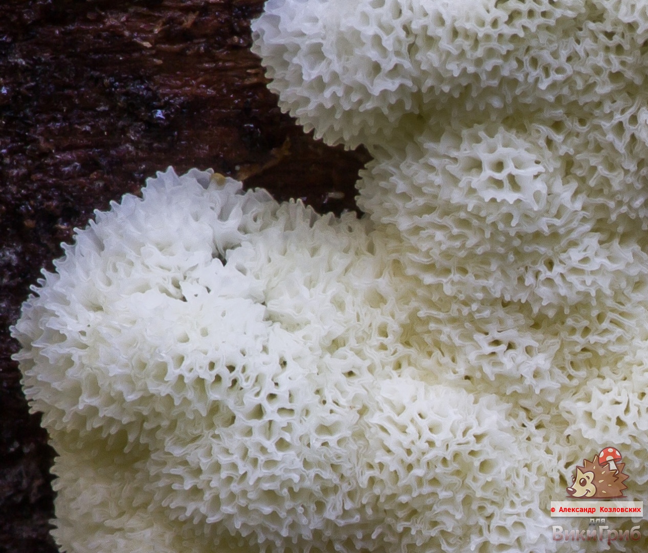
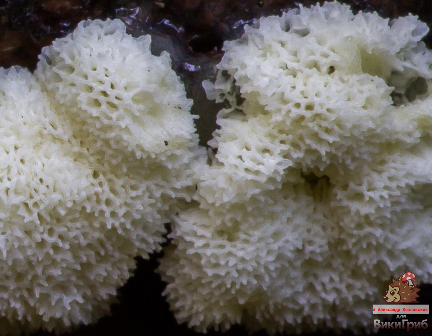
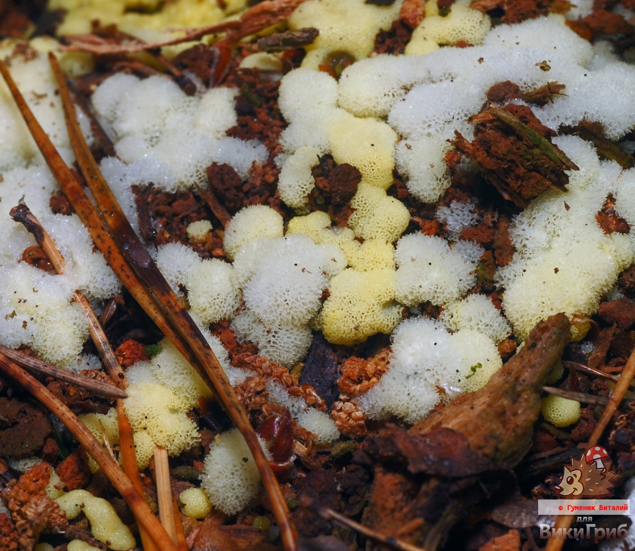
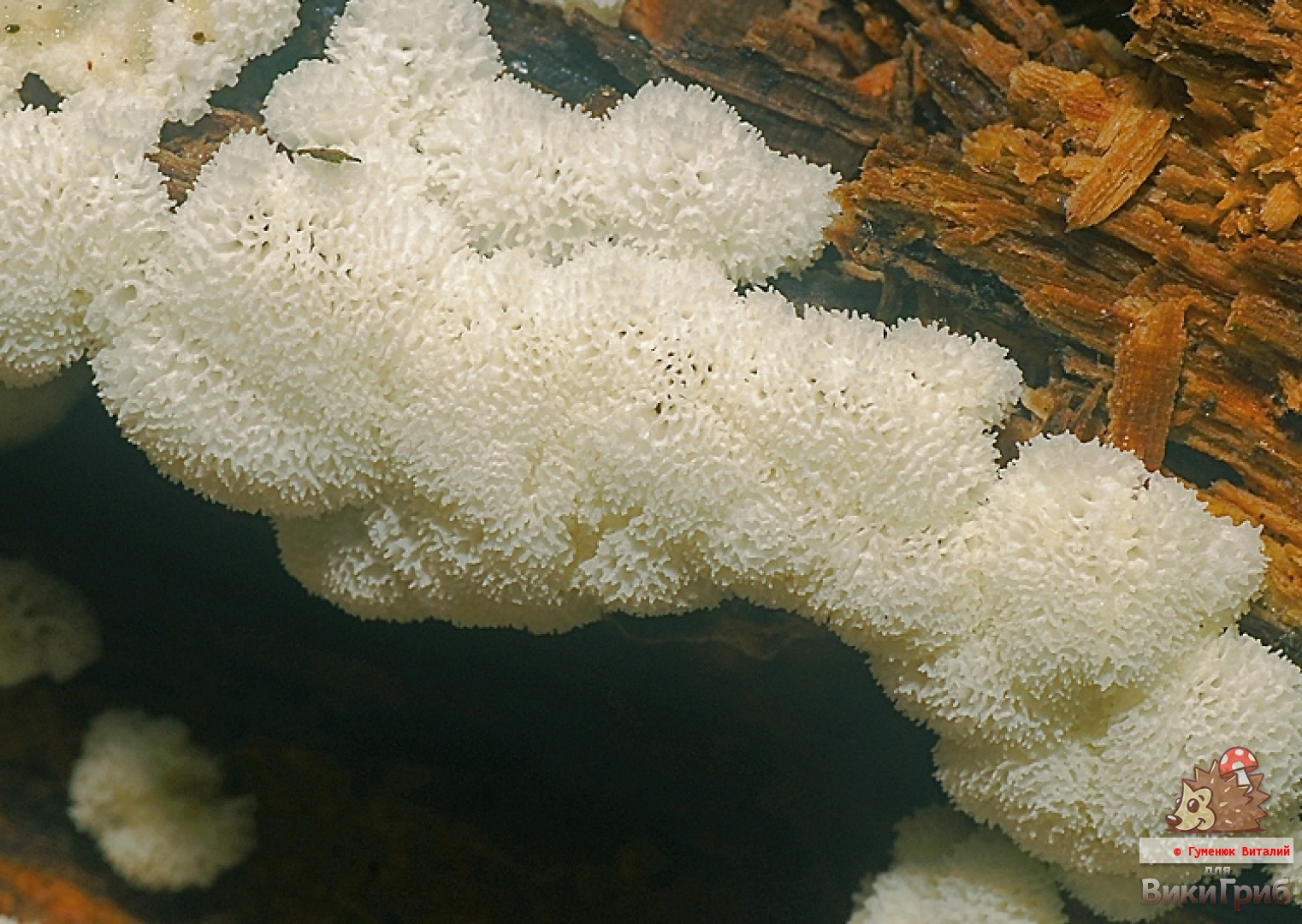
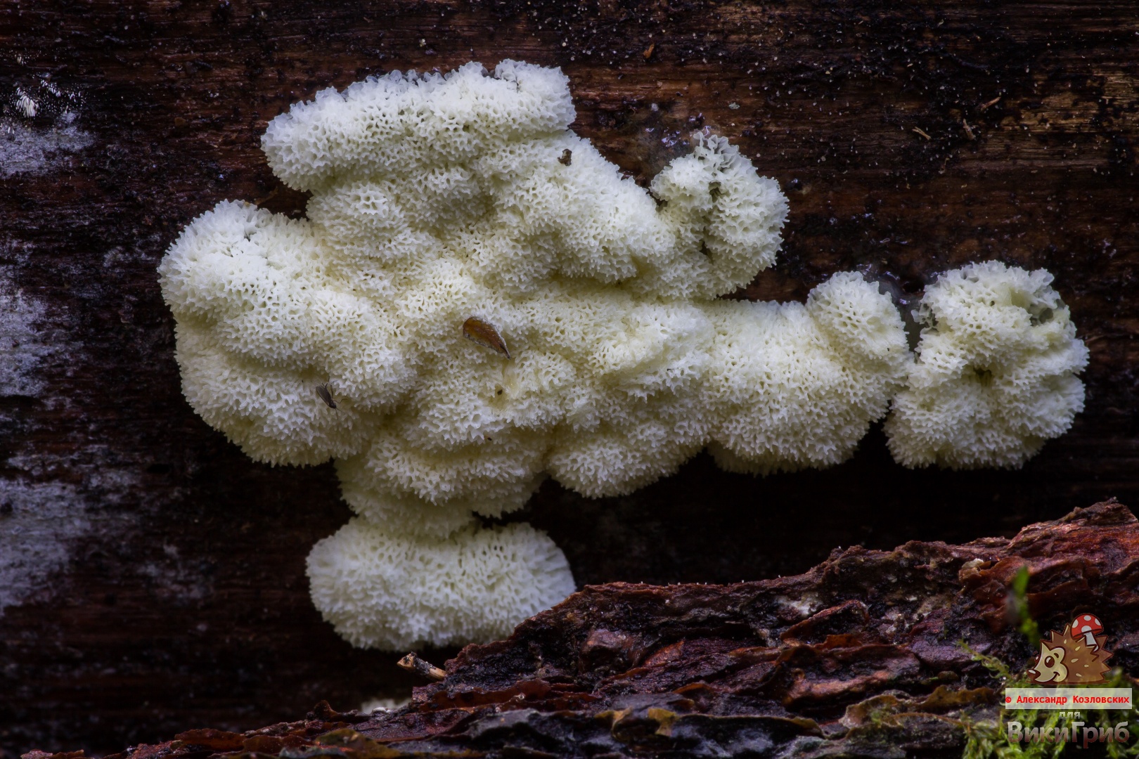
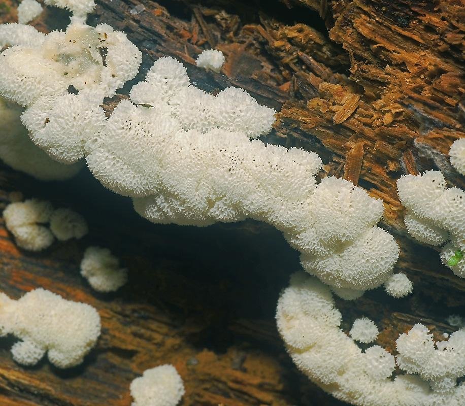
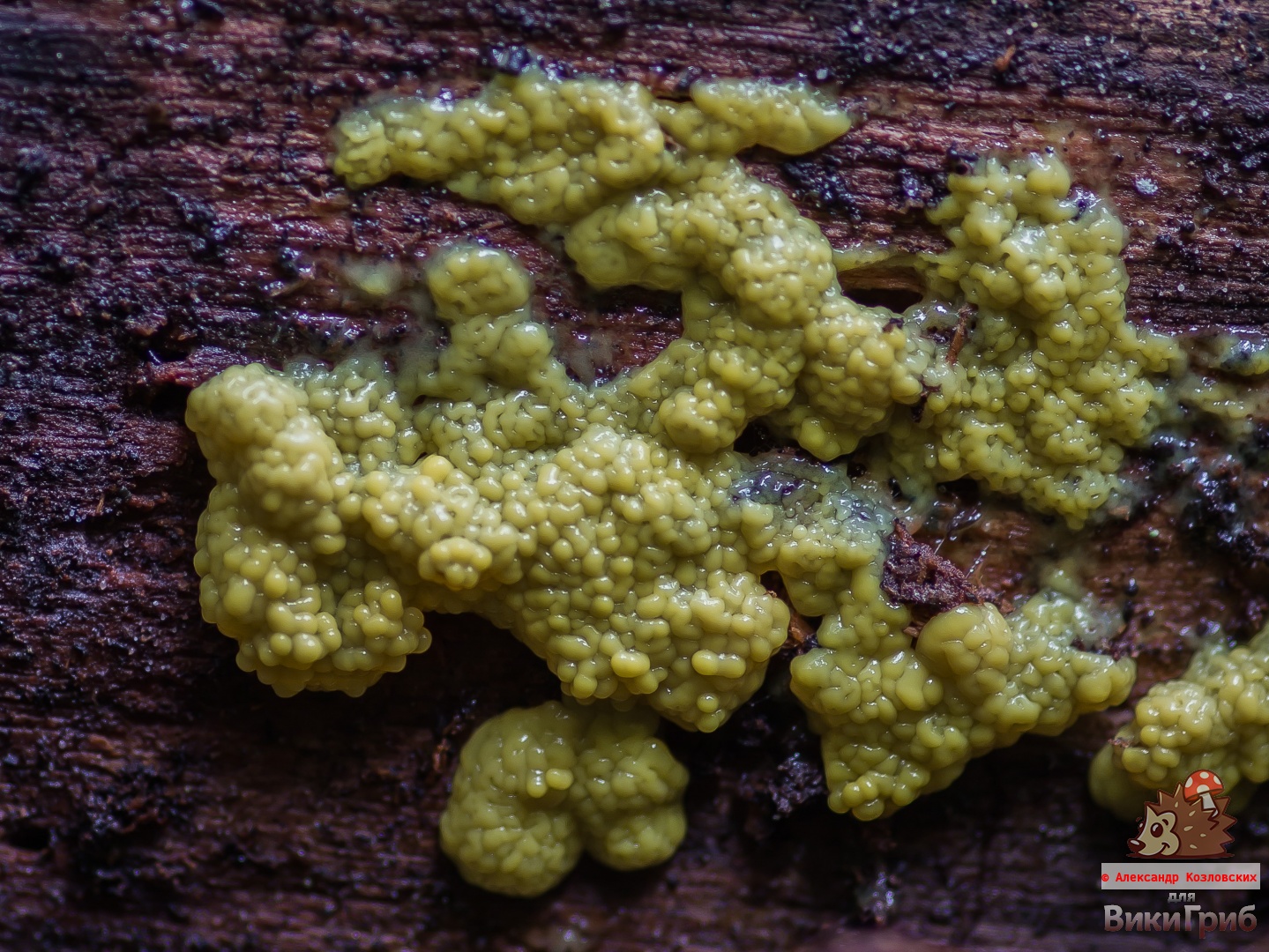
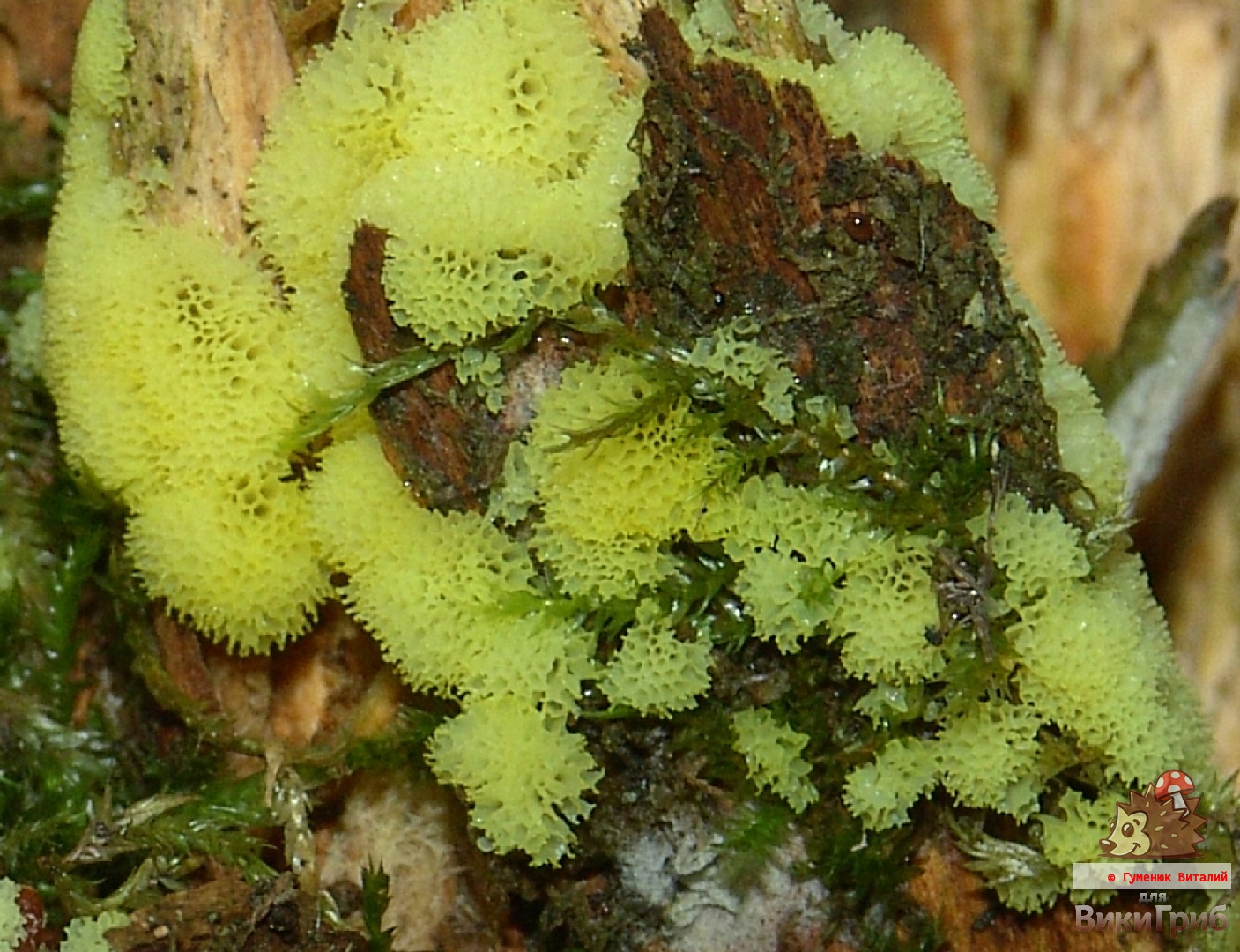
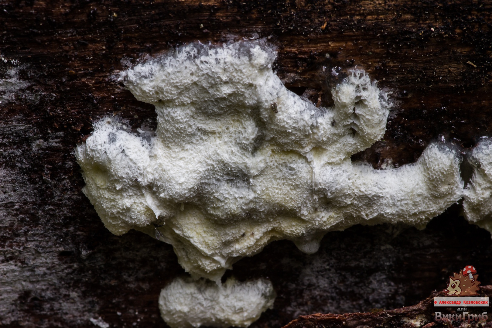
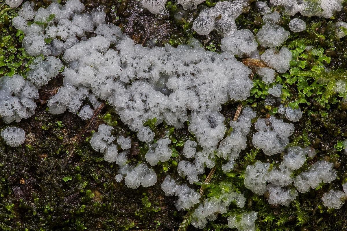
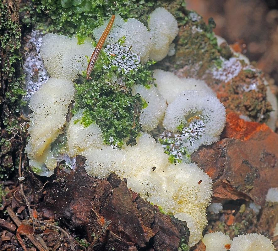
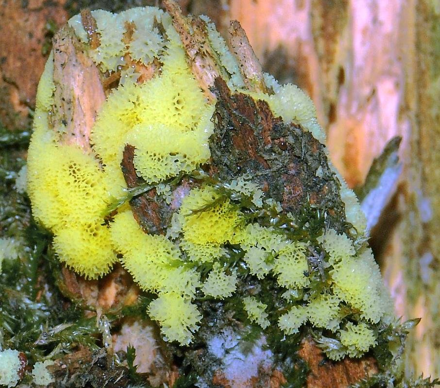
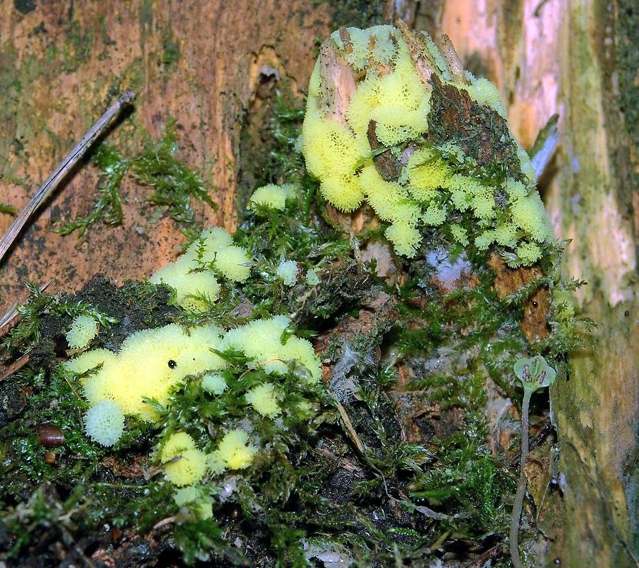
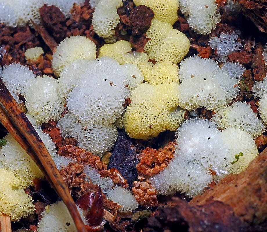
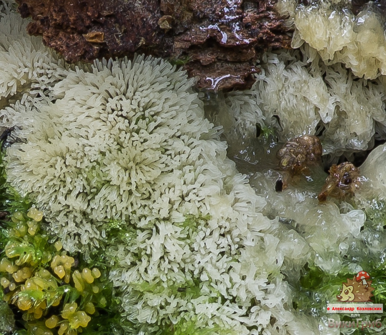
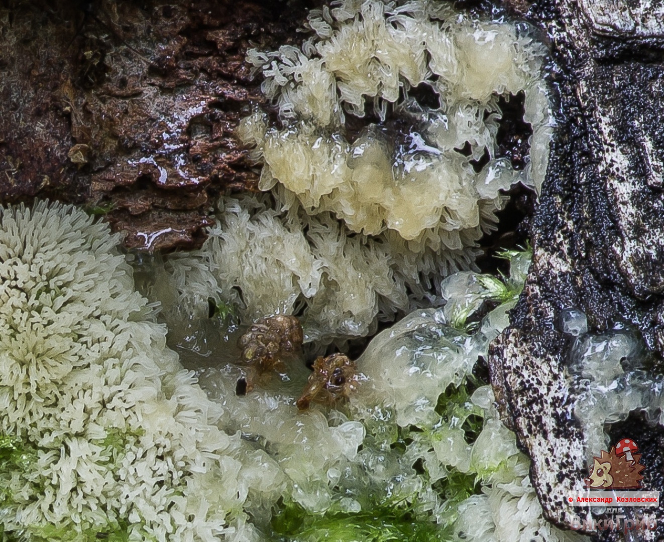
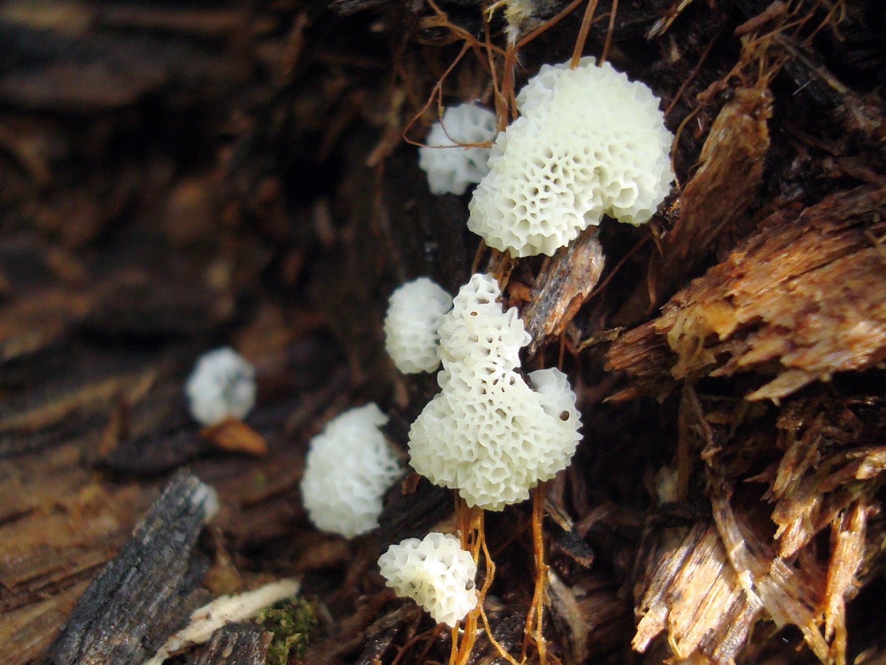
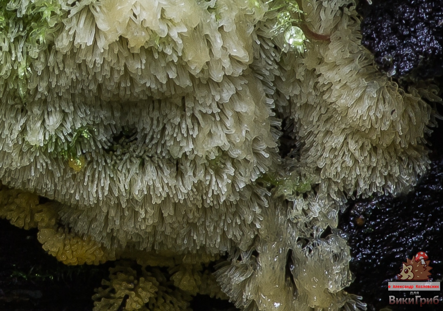
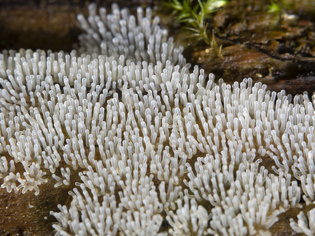
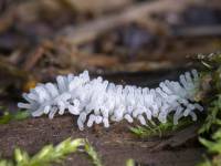
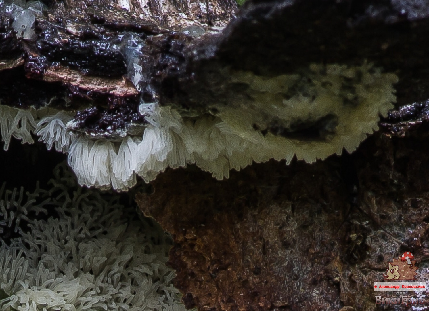
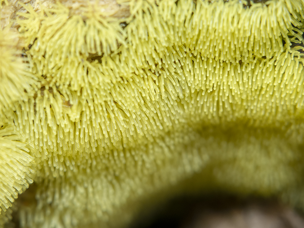
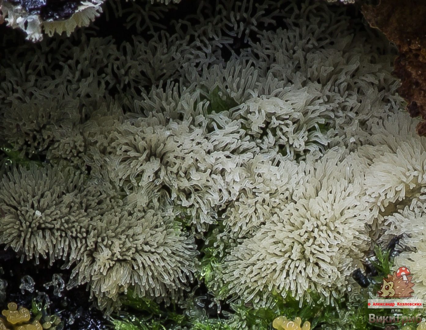
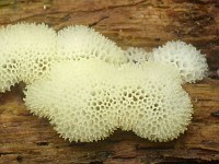
Unlike other myxomycetes, Ceratiomix dwarf shrub in the ripening stage consists of a number of vertical, simple or branched miniature columns, in the total mass taking the form of a porous, smooth or convex crust. White, but sometimes pinkish or pale yellow, yellowish greenish. It grows on average about 4 millimeters in height and forms extensive clusters on the surface of the substrate, covering an area from a few square centimeters to meters.
From a distance, to the naked eye, it looks like some kind of airy whitish glaze or a thin layer of foam. In order to see all the beauty of ceratiomixa, you need a magnifying glass or microshot.
Description
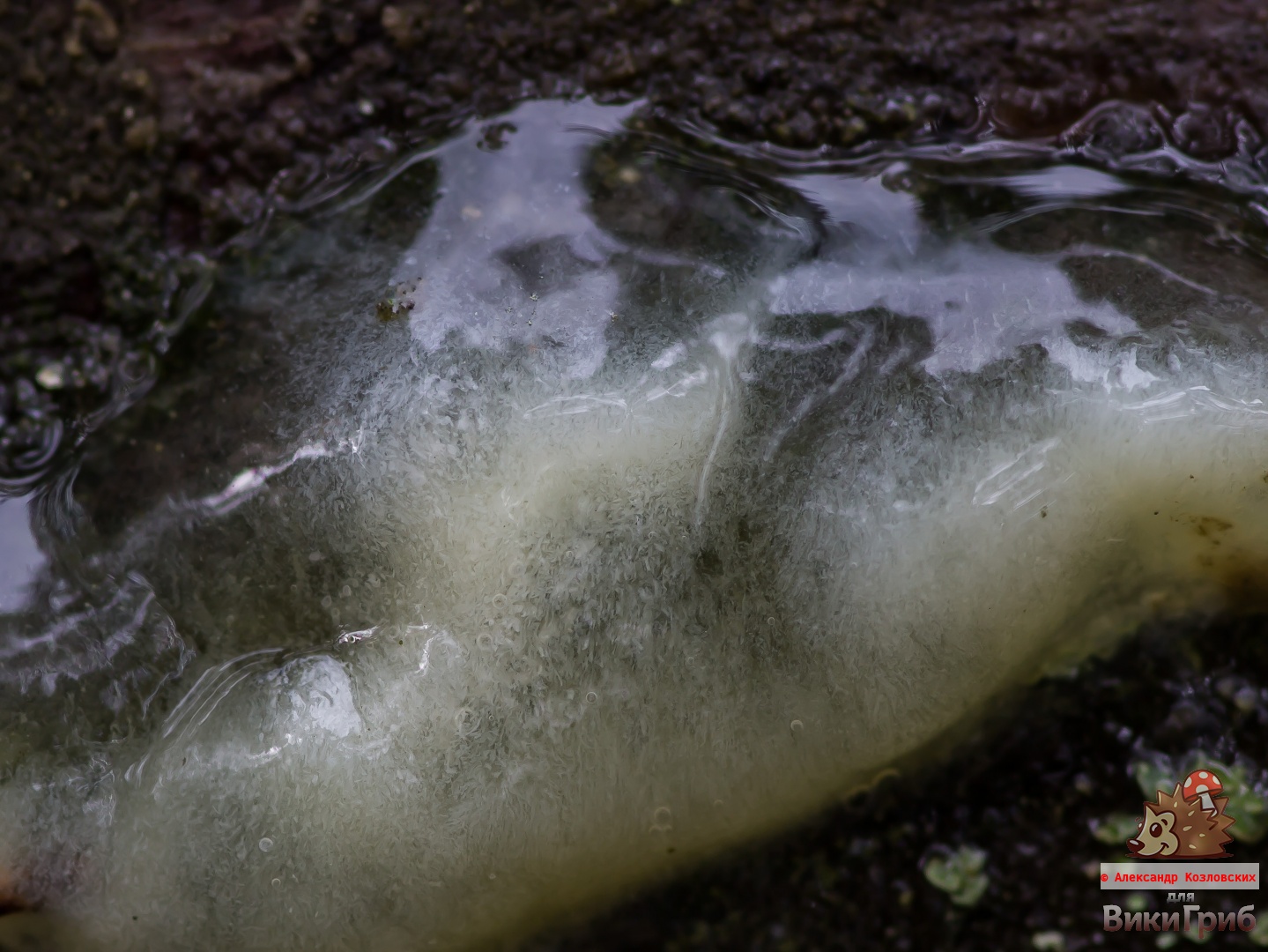
Plasmodium is white or yellowish.

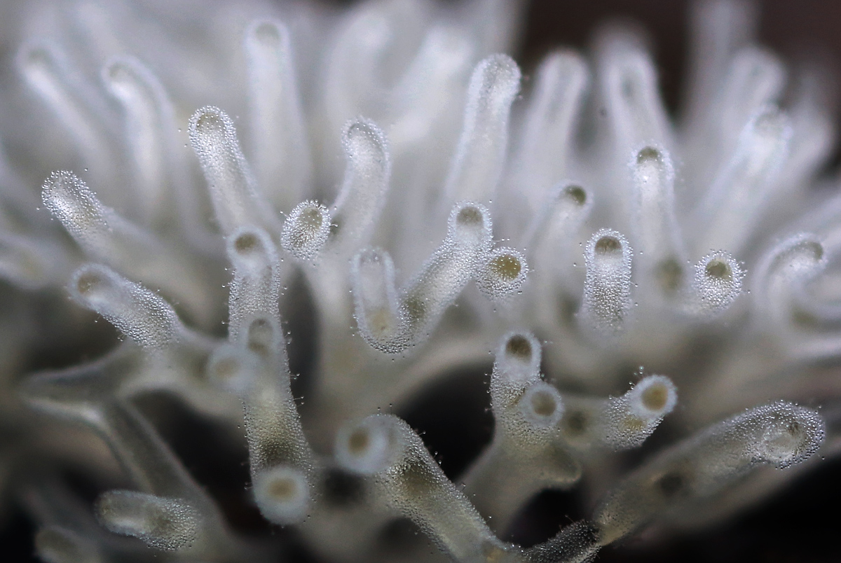
Sporocarps (fruiting bodies serving for the formation of spores) are very small. Height is approximately 1-6 (rarely up to 10) mm, thickness is 0.1-0.3, sometimes up to 0.5-1 mm. As a rule, they are white, transparent whitish, but there may be other colors, in yellowish, pinkish, yellowish-greenish or bluish tones. They look like tiny icicles.
Sporocarps in Ceratiomix are subshrub - columnar or coral-shaped, form simple or complex structures, sometimes branching near the base into several (up to 5) separate processes.
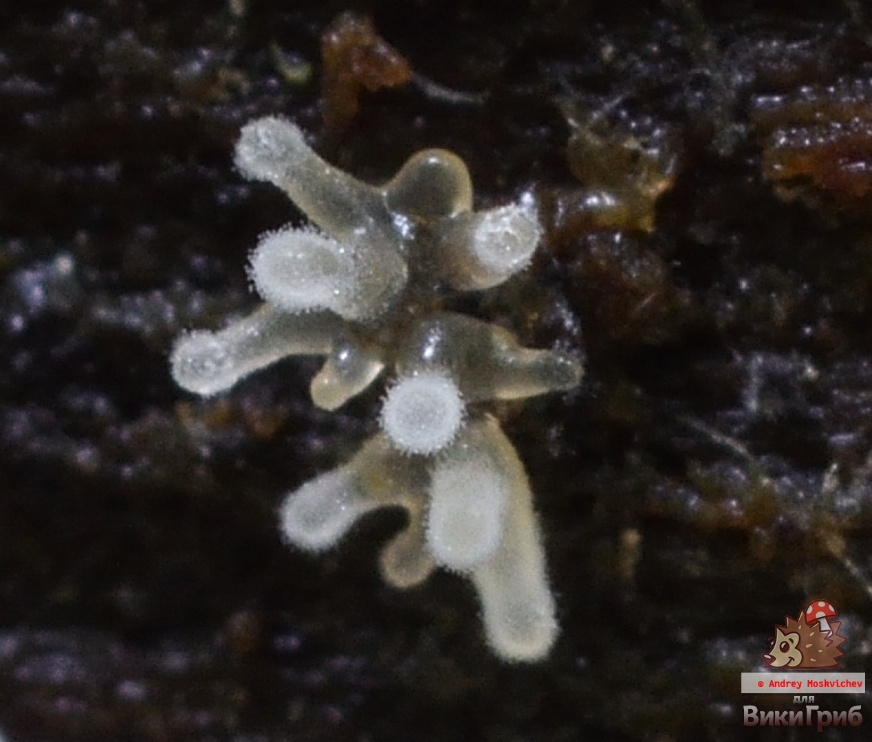
Individual sporocarps usually form more or less dense groups, in which dozens and hundreds of separate "columns" can be counted. This group has a soft, elastic texture.
Spores are formed on the outer surface of the sporocarps, therefore, in the photo, individual “branches” may have a slightly “blurred”, indistinct appearance.
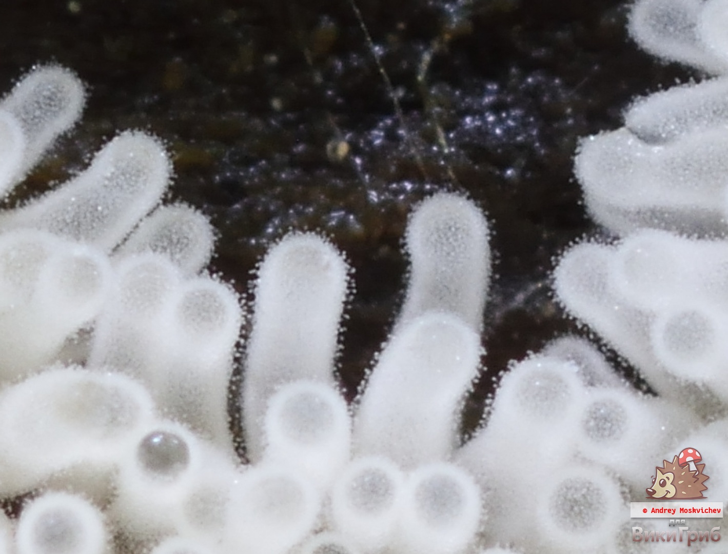

Colorless or pale greenish. The size of the spores is 7-20 x 1.5-3 microns.
Season and distribution
Cosmopolitan. Ceratiomix shrub is widespread in the tropics, and in the temperate zone, and in the Arctic.
It grows in the warm season, in summer and autumn, for the northern hemisphere, the dates are given: June-October, but adjustments made by weather conditions should be taken into account.
Ecology
Ceratiomix dwarf shrub grows on the surface of deciduous and coniferous trees and on mosses. Prefers dead wood, but can grow on the bark of living trees. This mixomycete does not parasitize on carriers and does not penetrate deeply into the organisms on which it grows. Plasmodium slowly moves along the surface of the substrate, absorbing particles of organic matter, bacteria and fungi.
Similar species
Other ceratiomixes. Other slime molds, of which there are a great many in nature, and not all of them are well described.
Subspecies of Ceratiomyxa fruticulosa:
- Ceratiomyxa fruticulosa f. aurantiaca
- Ceratiomyxa fruticulosa f. aurea
- Ceratiomyxa fruticulosa f. flava
- Ceratiomyxa fruticulosa f. fruticulosa
- Ceratiomyxa fruticulosa f. rosea
- Ceratiomyxa fruticulosa var. arbuscula
- Ceratiomyxa fruticulosa var. caesia
- Ceratiomyxa fruticulosa var. comata
- Ceratiomyxa fruticulosa var. descendens
- Ceratiomyxa fruticulosa var. flexuosa
- Ceratiomyxa fruticulosa var. fruticulosa
- Ceratiomyxa fruticulosa var. porioides
- Ceratiomyxa fruticulosa var. rosella
Photo: Vitaly Gumenyuk, Alexander Kozlovskikh, Andrey Moskvichyov.
Who are slime molds
Slime molds (eng. slime molds) - mobile terrestrial unicellular phagotrophs, forming large spore-bearing structures, fruiting bodies... Their macroscopic size and terrestrial lifestyle put them on a par with four other groups: multicellular animals, green plants, real fungi, and supercolonial cyanobacteria (for example, Nostoc pruniforme). But among the members of this "club of heavyweights" slime molds stand out for the complete absence of true multicellularity, that is, they do not form a mass of physiologically related cells originating from a single progenitor cell. They have neither tissues, nor organs, nor even their analogs, as in higher fungi. The slime mold begins life in the form of a microscopic amoeba, which, after a series of dramatic transformations, turns into a scattering of large and brightly colored spore-receptacles, which have a different, but in any case, not a tissue structure.
Scientists and ordinary people for many centuries have taken the spore-bearing structures of slime molds for the fruiting bodies of mushrooms. The situation changed in the 1820s, after the eminent mycologist Elias Magnus Freese forgot his hat-cylinder in the forest, in which he put the immature fruit body of a slime mold in the forest for safekeeping. Returning for his hat in the evening, Freese discovered that a strange mushroom ... had crawled out onto the brim of the cylinder, where it finally froze and matured. What seemed to be the fruiting body of the mycelium hidden in the thickness of the earth was the sporulation of a giant mobile creature! When Anton de Bary in 1858 germinated slime mold spores in the laboratory and discovered that tiny amoebas were crawling out of them, it became clear that slime molds have no more to do with fungi than whales have with fish.
The fruiting bodies of slime molds can be formed in two different ways. Way aggregations suggests that free cells, resembling ordinary amoebas, gather in close groups, the so-called pseudoplasmodium (Fig. 2, a, b). A mature pseudoplasmodium behaves like a single multicellular organism: it maintains a constant shape, moves purposefully along the substrate, and finally, as a result of a rather complex "embryogenesis", forms a "ball on a leg" structure - the fruiting body itself. Pseudoplasmodia and fruiting bodies do not develop from a single progenitor cell, like the bodies of multicellular organisms, but are temporary associations of genetically diverse individuals. At the same time, in the course of ontogenesis of fruiting bodies, some amoeba can be purposefully sacrificed, since the leg, consisting of dead cells, is stronger than the "living" one.Such a strategy - American biologist James Cavender jokingly called it "communist" - brings its owners closer to truly multicellular organisms. Slime molds, which are characterized by aggregation and pseudoplasmodia, are traditionally called cellular.
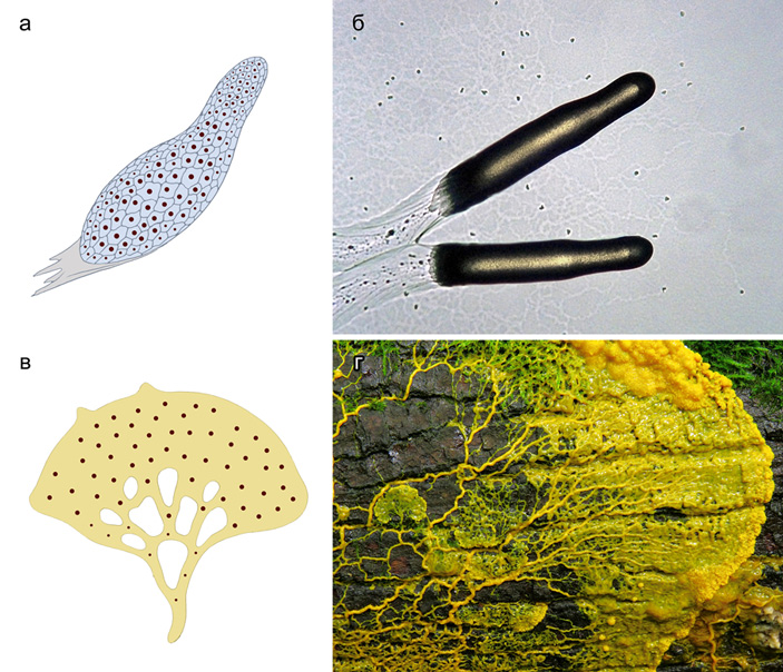
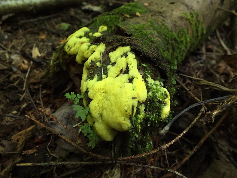
The second way of development, plasmodation, allows you to get a complex fruiting body from a single microscopic amoeba. True, for this it has to become macroscopic, expanding, but not dividing. As a result, plasmodium (Fig. 2, c, d), a giant multinucleated amoeba that can cover an area of up to 1 m2, be up to 5 m in length and weigh up to 20 kg (in a slime mold Brefeldia maxima). Most Plasmodia, of course, are not so huge, but large enough to be eaten in Latin America. In this case, the plasmodium is typical, having, however, millions of nuclei, an extremely powerful cytoskeleton and a complex cytoplasmic circulation system. Having reached a certain size, the plasmodium proceeds to the formation of fruit chalk. Being single-celled, it cannot sacrifice some of its cells to build auxiliary structures. Therefore, all components of the fruiting body, except for spores, are formed from deposits of polysaccharides (mainly β-1,4-galactosaminoglycan, see glycosaminoglycans), which accumulate on the surface of the plasmodium or inside the cisterns of its endoplasmic reticulum. In some species, a small number of spore-like cells may remain in the stem, but this is more an accident than a deliberate strategy. Slime molds, which are characterized by plasmodation, are usually called plasmodial.

