Definitioner
- Basidia (Basidia)
-
Lat. Basidia. A specialized structure of sexual reproduction in fungi, inherent only in Basidiomycetes. Basidia are terminal (end) elements of hyphae of various shapes and sizes, on which spores develop exogenously (outside).
Basidia are diverse in structure and method of attachment to hyphae.
According to the position relative to the axis of the hypha, to which they are attached, three types of basidia are distinguished:
Apical basidia are formed from the terminal cell of the hypha and are located parallel to its axis.
Pleurobasidia are formed from lateral processes and are located perpendicular to the axis of the hypha, which continues to grow and can form new processes with basidia.
Subasidia are formed from a lateral process, turned perpendicular to the axis of the hypha, which, after the formation of one basidium, stops its growth.
Based on morphology:
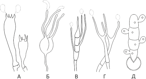
Holobasidia - unicellular basidia, not divided by septa (see Fig. A, D.).
Phragmobasidia are divided by transverse or vertical septa, usually into four cells (see Fig. B, C).
By type of development:
Heterobasidia consists of two parts - hypobasidia and epibasidia developing from it, with or without partitions (see Fig. C, B) (see Fig. D).
Homobasidia is not divided into hypo- and epibasidia and in all cases is considered holobasidia (Fig. A).
Basidia is the place of karyogamy, meiosis and the formation of basidiospores. Homobasidia, as a rule, is not functionally divided, and meiosis follows karyogamy in it. However, basidia can be divided into probasidia - the site of karyogamy and metabasidia - the site of meiosis. Probasidium is often a dormant spore, for example in rust fungi. In such cases, probazidia grows with metabasidia, in which meiosis occurs and on which basidiospores are formed (see Fig. E).
See Karyogamy, Meiosis, Gifa.
- Pileipellis
-
Lat. Pileipellis, skin - differentiated surface layer of the cap of agaricoid basidiomycetes. The structure of the skin in most cases differs from the inner flesh of the cap and may have a different structure. The structural features of pileipellis are often used as diagnostic features in descriptions of fungi species.
According to their structure, they are divided into four main types: cutis, trichoderma, hymeniderma and epithelium.
See Agaricoid fungi, Basidiomycete, Cutis, Trichoderma, Gimeniderm, Epithelium.
Syzygospore mushroom-loving, Syzygospora mycetophila
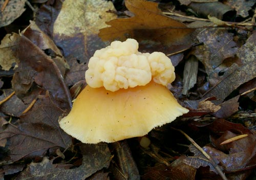
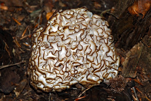
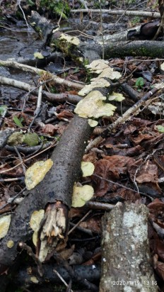
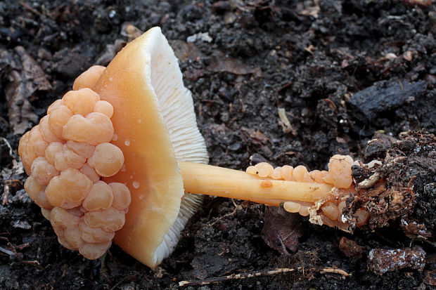
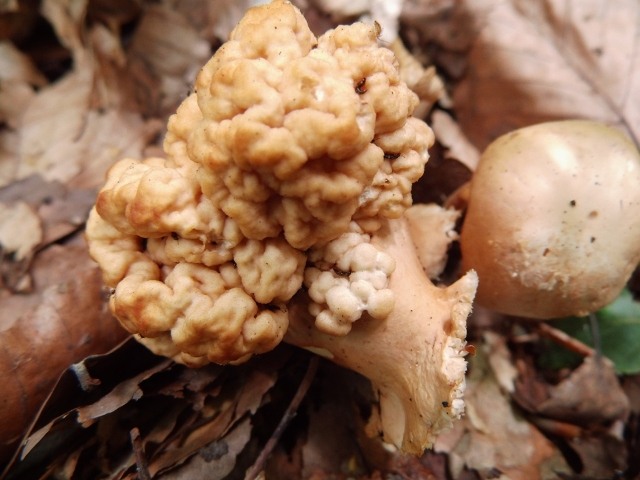
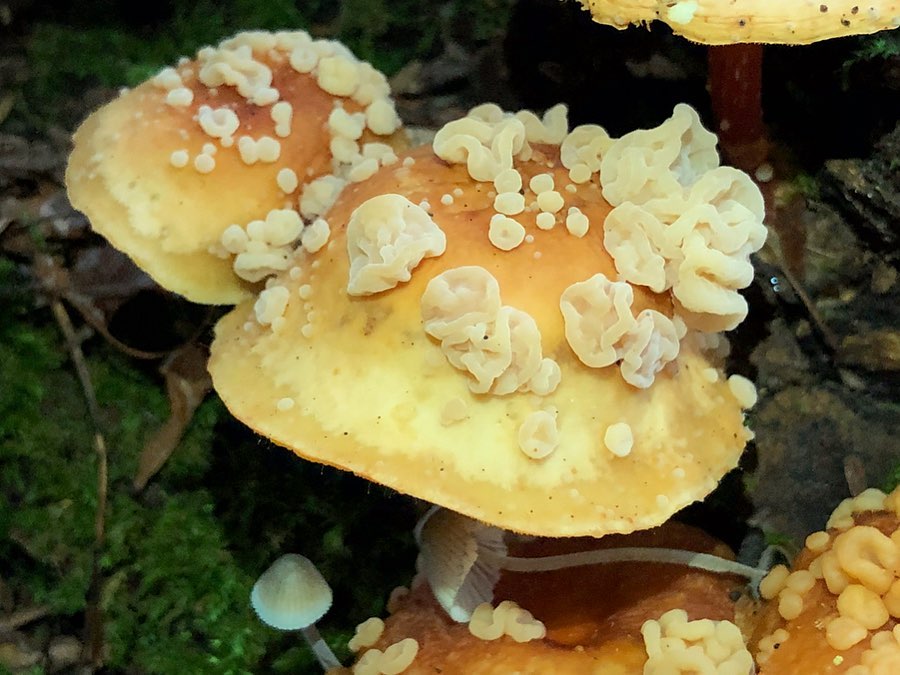
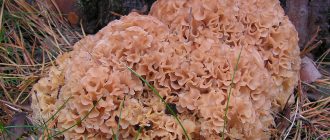
Fruit body: Sedentary, cerebral, yellowish-brown in color. In the process of development, galls 0.5 - 1 cm in size formed on the cap (less often at the base of the leg) of the host fungus merge together, forming one giant brain, which used to completely cover the fruiting body of the host fungus. The pulp has an elastic, slightly "gelatinous" consistency, reminding us of the relationship of the mushroom with "shivers".
Spore Powder: Apparently white.
Distribution: Syzygospora mycetophila parasitizes on the fruiting bodies of the tree-loving Collybia dryophila and the oily C. butyracea, developing, as can be judged from observation experience, mainly in summer, in hot and humid weather. It occurs infrequently, however, under appropriate conditions, a significant number of fungi over a large area can be infected with tree-loving syzygospore.
Similar species: It must be recognized that the definition of the fungi shown here as Syzygospora mycetophila is not entirely serious. The fruiting bodies of the syzygospore are very variable, depending on the growing conditions, the state of the host fungus and their own age, therefore, one should not rely on their appearance alone for an accurate determination. Syzygospora mycetophila can only be distinguished from S. tumefaciens or S. effibulata by measuring the spores. However, since the mushroom-loving syzygospore is found in our area more often than its relatives (at least this conclusion can be drawn from the fragmentary information presented on the Internet), we will not multiply the entities and call the find as it is most likely.
Edible: It hardly crossed anyone's mind.
Author's Notes: The brain-like mushroom has all sorts of jokes about itself, as well as about the person who found it and everyone to whom it managed to tell about it. However, jokes are jokes, however, a certain symbolism is visible in this syzygospore, either gloomy, or life-affirming - depending on what position to consider what is happening. Judge for yourself: a small, inconspicuous and completely uninteresting mushroom, a simple colibia, suddenly attacks and masters something of a very intelligent look, often leaving not the slightest trace of the unfortunate owner. How can we not recall the alarming esoteric concepts, within which the mind is a “rider” of a person, an alien installation that pursues its own goals and makes a person think that he really wants to do everything that people usually do?
However, down with doubts - if the parasites of consciousness existed, they would have been shown on TV long ago (although where is the guarantee that they are not shown?). Better still about mushrooms. Now I know very well where to get this syzygospore according to the season, and I can quite close the question of its species belonging even before the end of the coming summer, however, to my horror, I do not understand where the spore-bearing layer is located in this mushroom. Outside? This is not some line. Inside? How then do spores get to the surface, looking for new victims? After all, then, outside? Or does Syzygospora mycetophila besides “inside” and “outside” have some other geometric quality, hitherto unknown? We didn't even have time to start, but already how many obstacles, how many unanswered questions and unverifiable guesses.
Well, before the start of the season there is still time to get the brain out of my head and again be puzzled by the problem, suddenly stumbling upon a suspicious brain structure in a familiar forest ...

