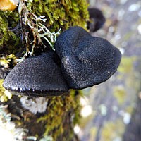
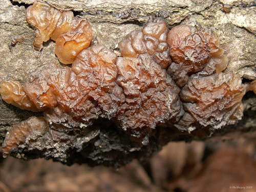
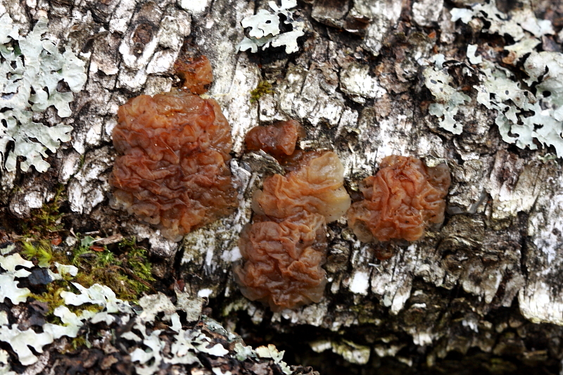
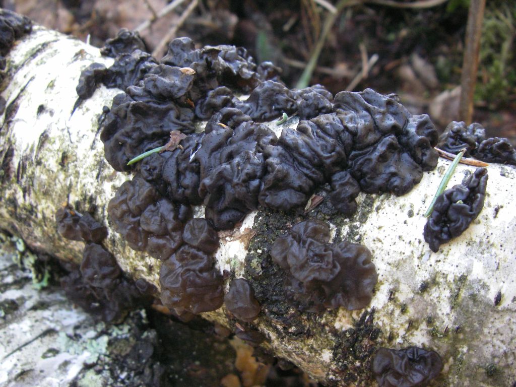
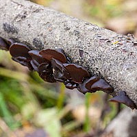
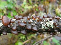
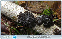
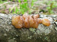
Genus: Exidia (Ex> Main Fungi Bas> Exidia
Exidia is a genus of fungi of the Auriculariaceae family.
Etymology
Exidia, Exidia. From lat. exsudo, avi, atum, are, go out, stand out.
Typical view
Description
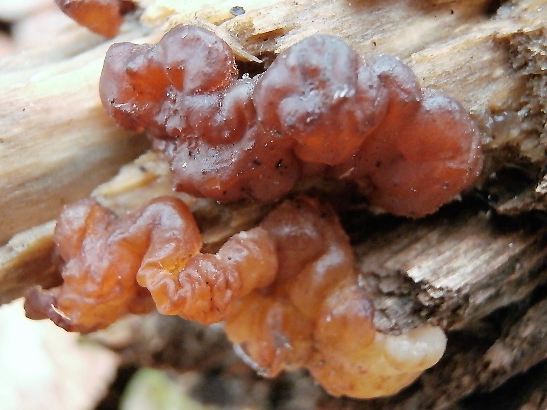
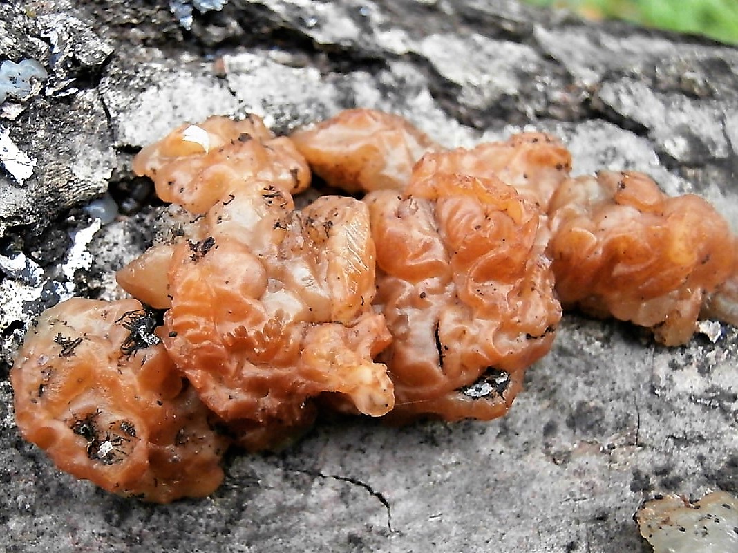
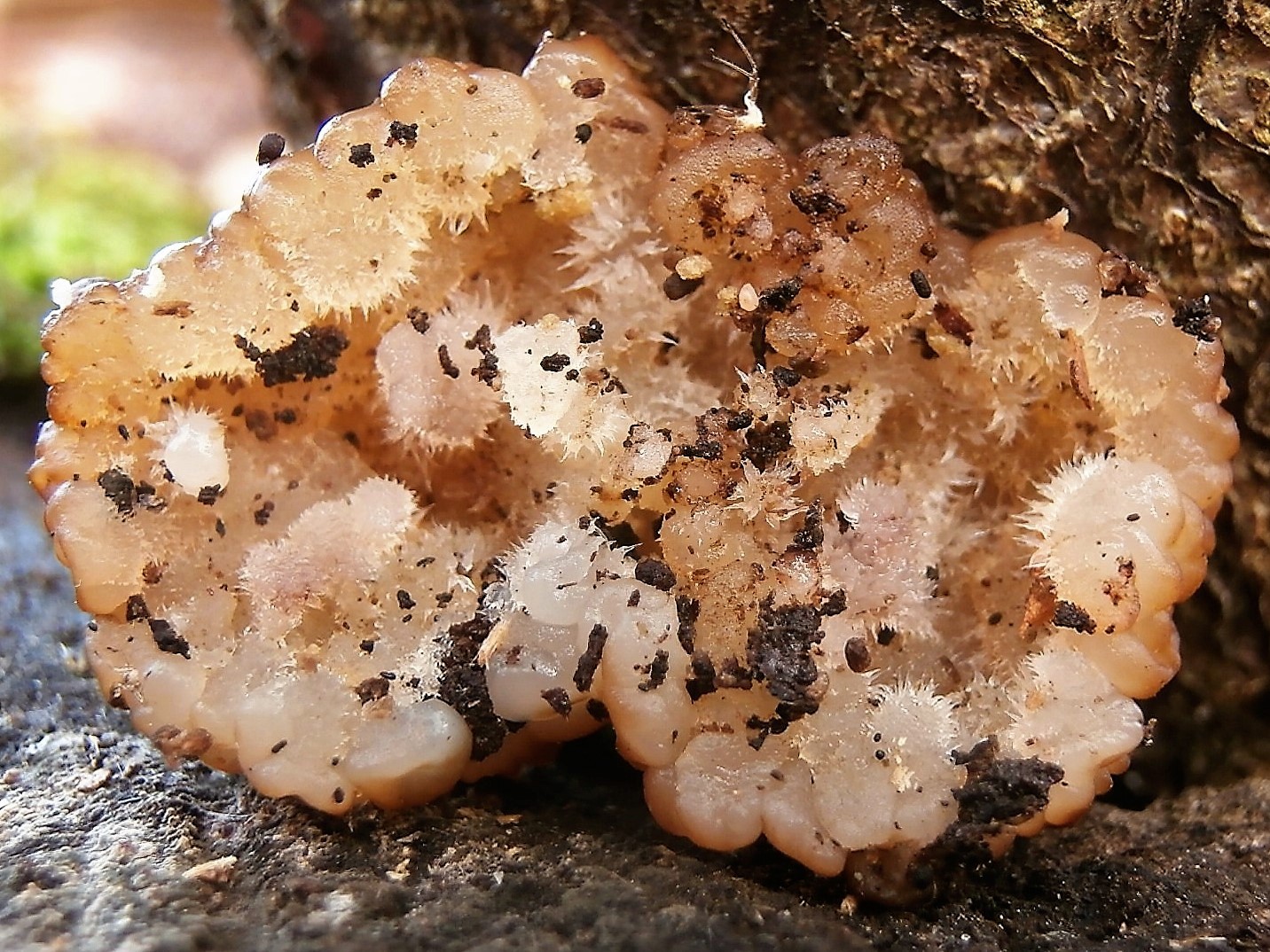
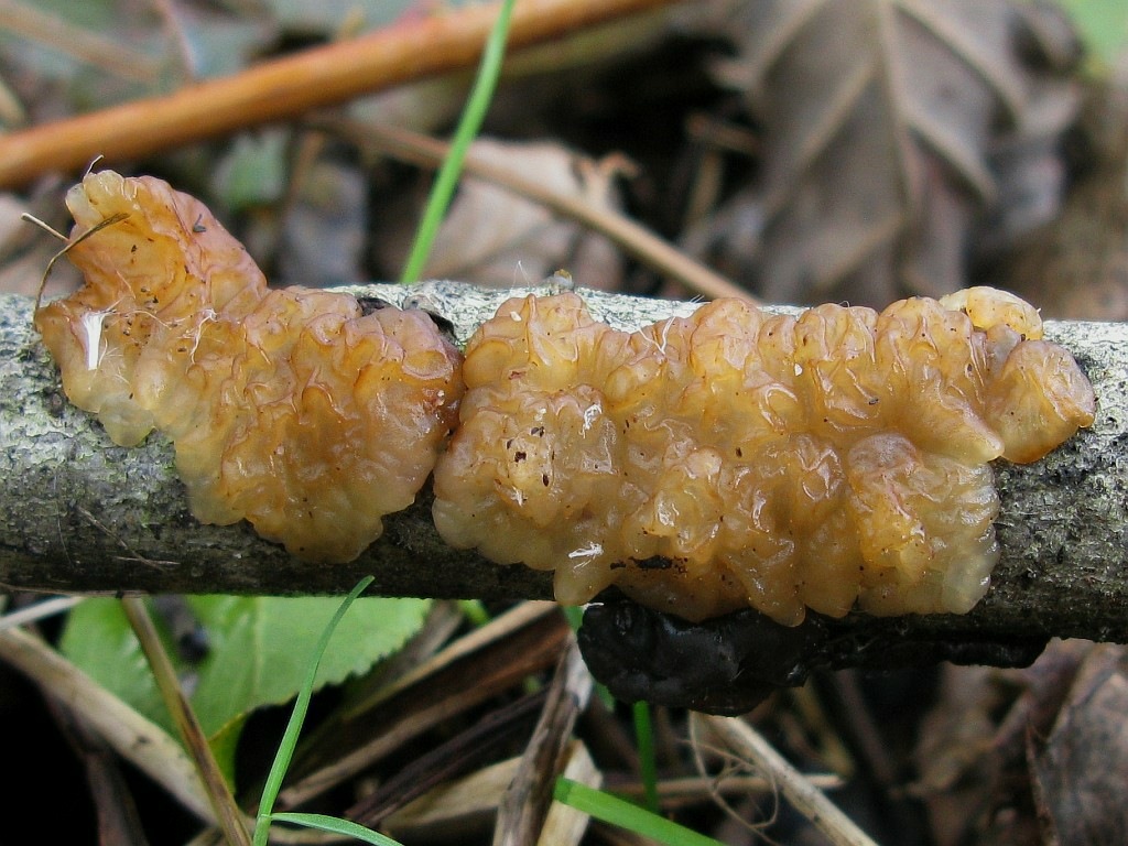
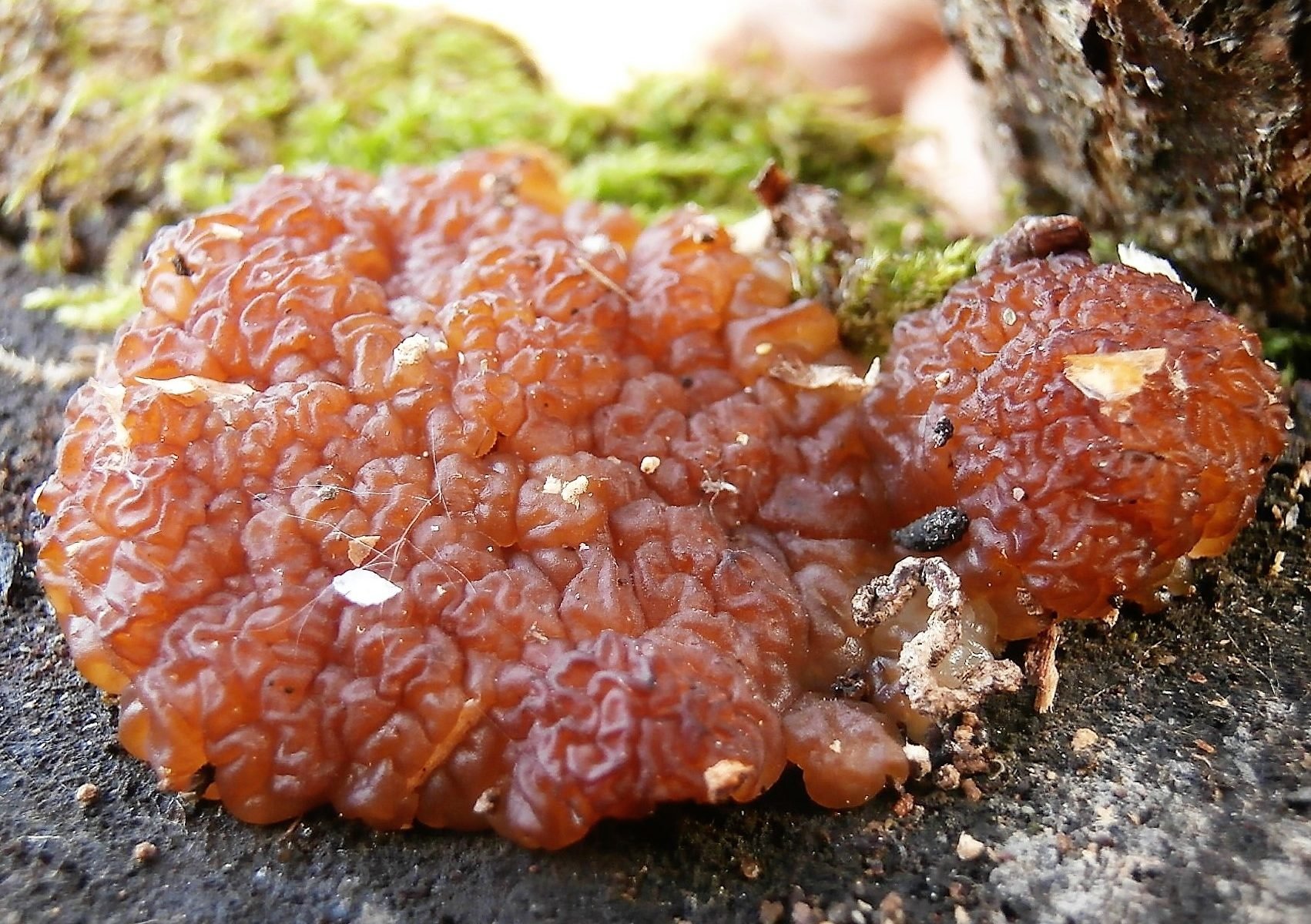
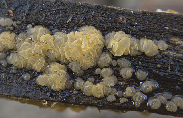
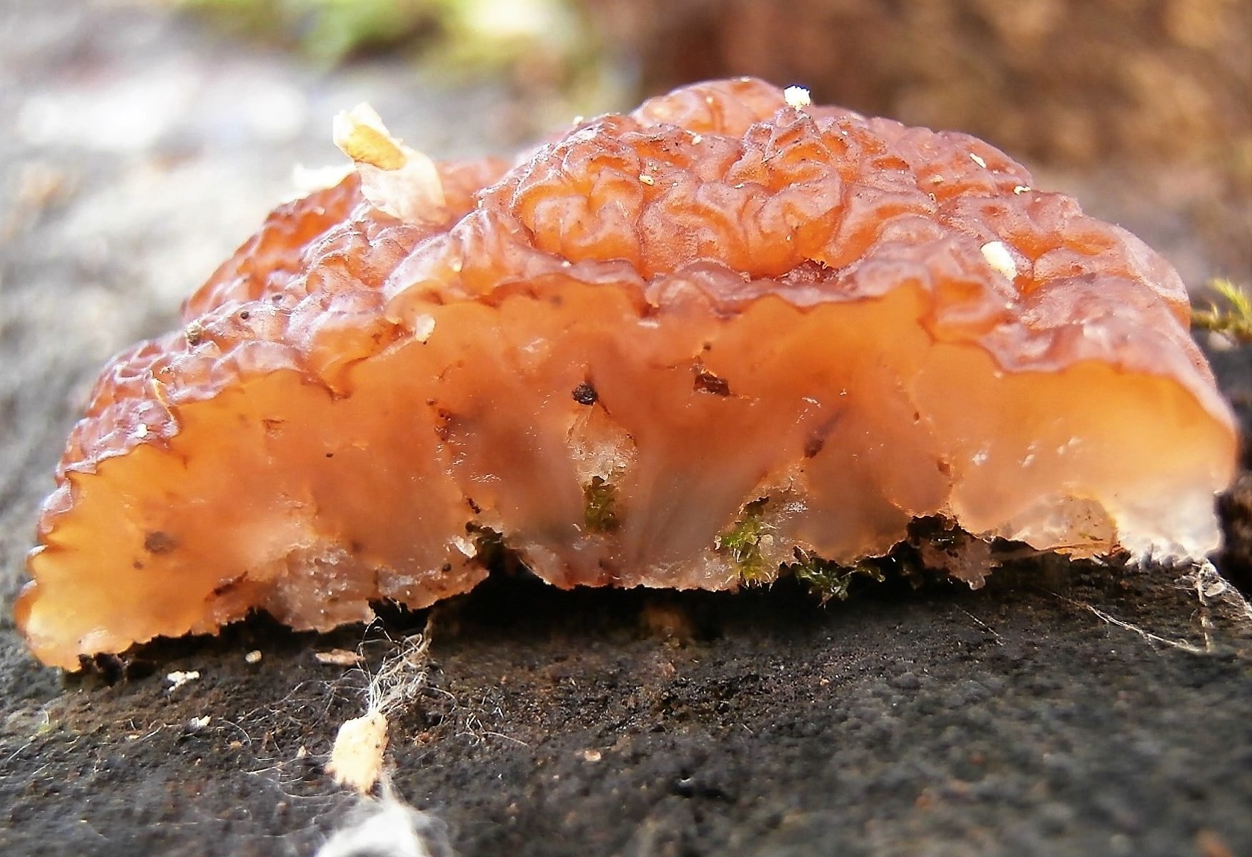
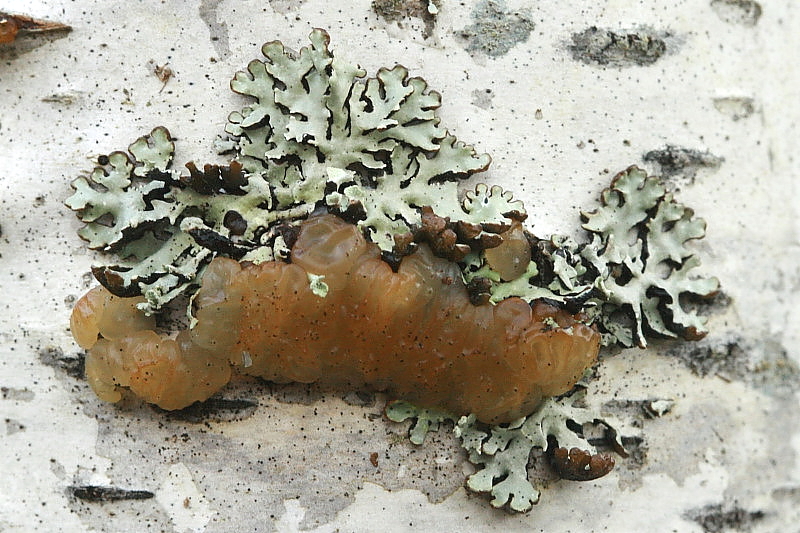
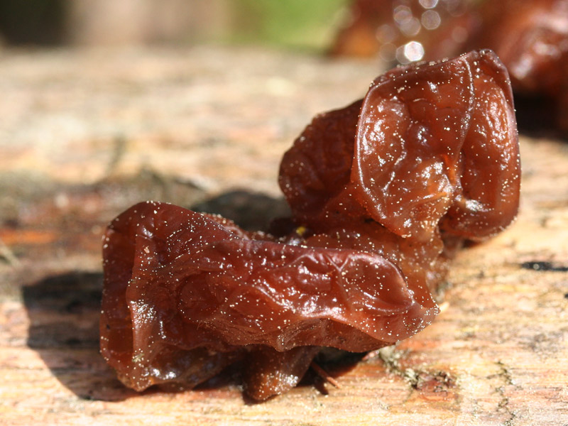
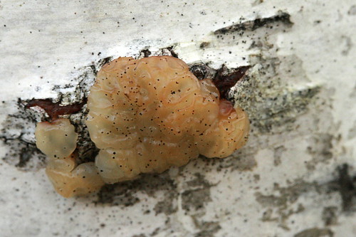
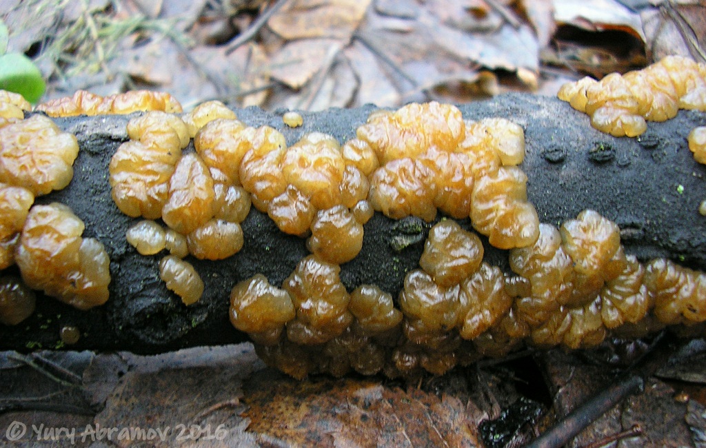
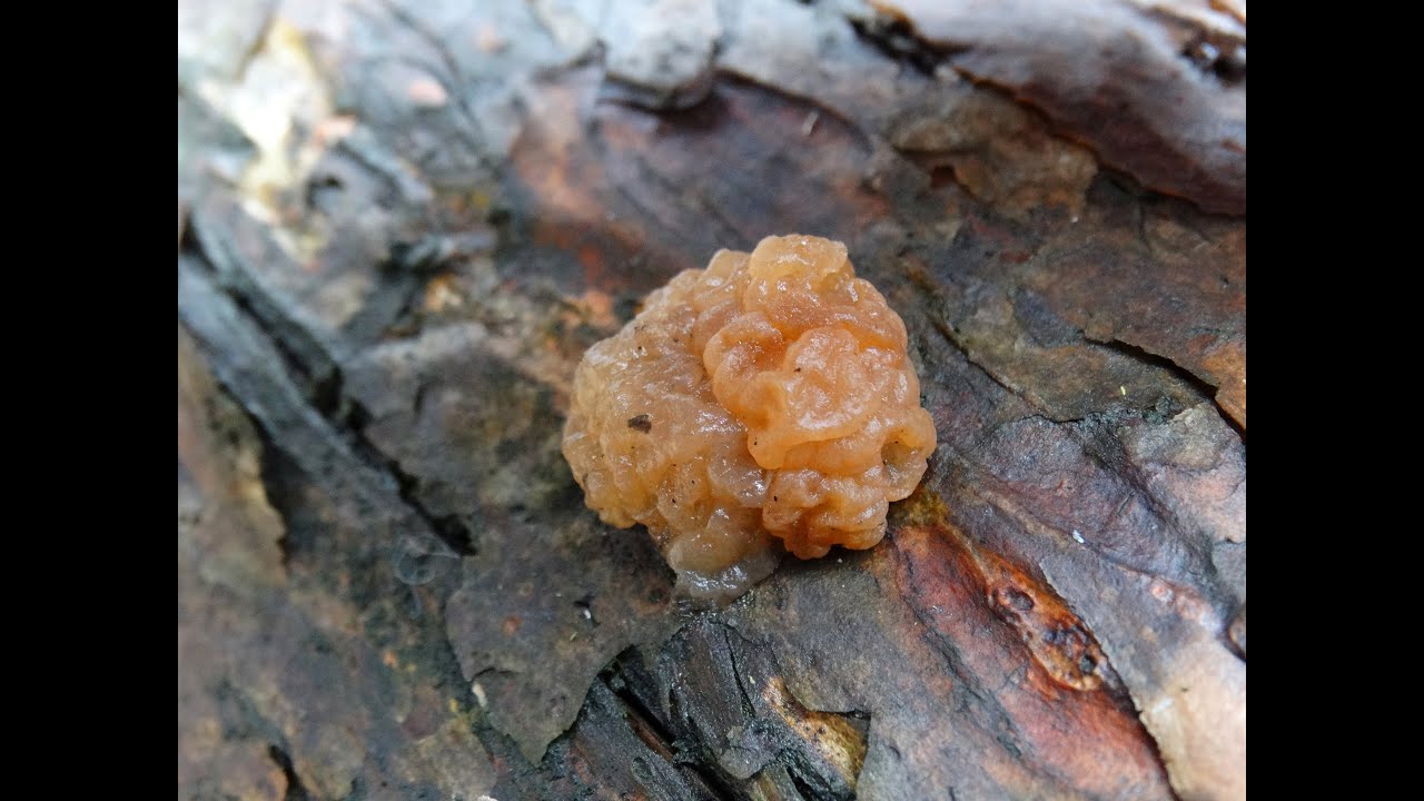
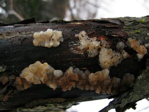
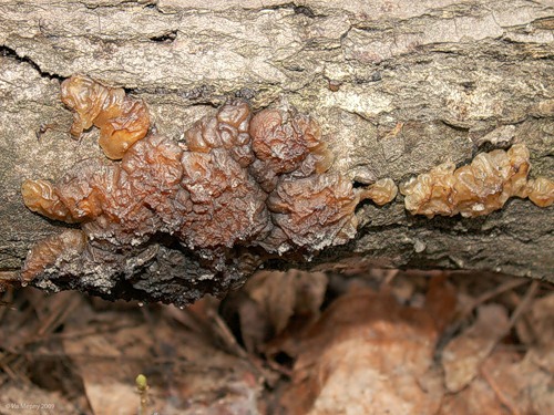
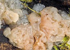
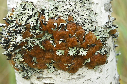
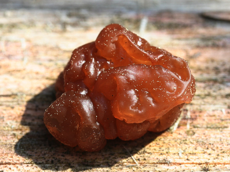
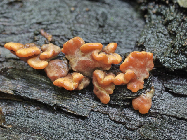
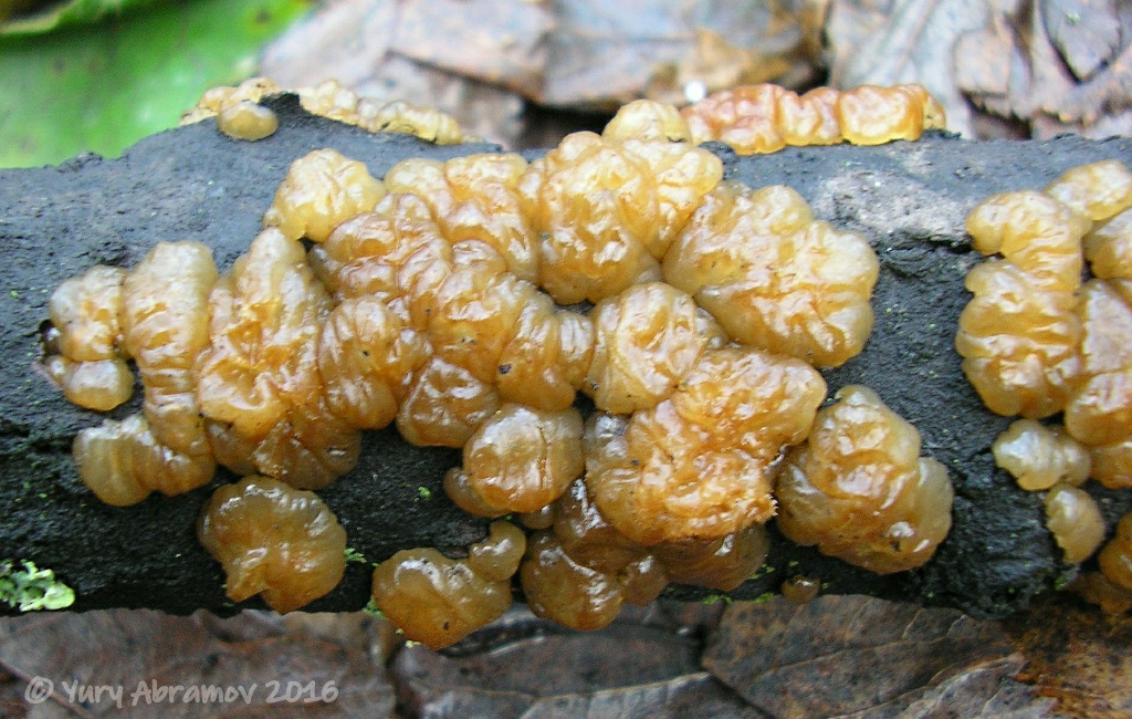
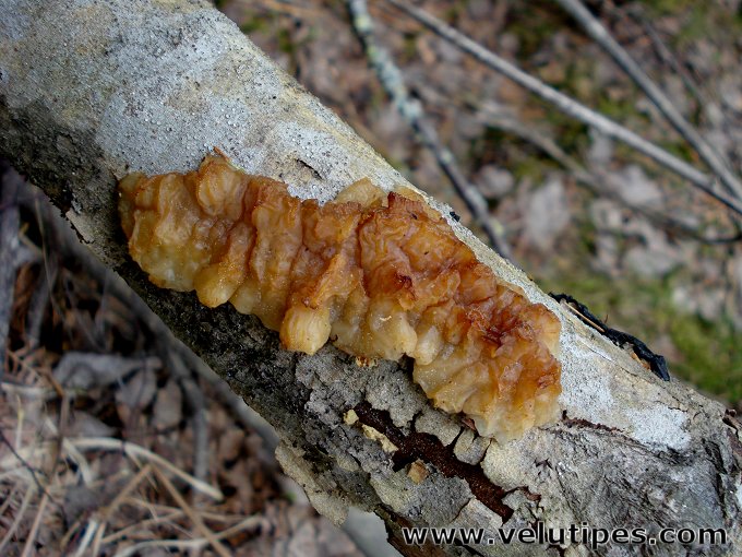
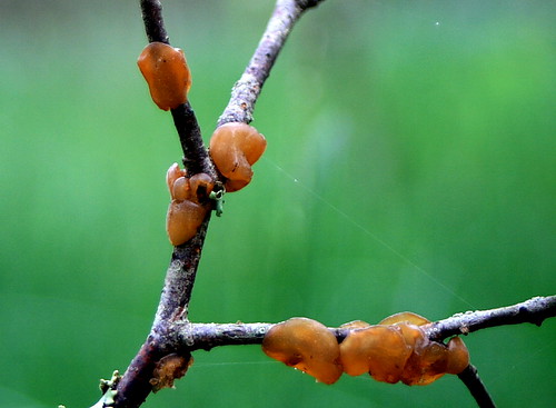
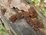
Fruit bodies are varied in shape (from cushion, disc-shaped and fusiform to sinuous, lobed and prostrate), as well as varied in color (from almost colorless, white and yellowish to various shades of brown and black). The surface of the fruit bodies is smooth, wrinkled, or covered with tubercles. The consistency of the fruiting bodies is soft or densely gelatinous or cartilaginous. The hymenium is covered with an epigymenal membrane. Fabric hyphae with buckles. Basidia are almost spherical, ovate or ellipsoid, 4-spore, cruciform-septate. Spores are smooth, hyaline, allantoid.
They grow on deciduous and coniferous wood.
Key for identifying species of the genus Exidia distributed in Russia
1. Fruit bodies are black → 2.
- Fruit bodies of a different color → 4.
2. On conifers → Exidia pithya.
- On hardwoods → 3.
3. Fruit bodies are cushion-shaped, irregular in shape, merging into a single mass with the loss of isolation → Exidia nigricans.
- Fruit bodies are compact, discrete, which, even if they form groups, never merge into a single mass with the loss of isolation of individual fruit chalk. Occurs on broadleaf species → Exidia glandulosa.
4. Fruit bodies are hyaline, white or yellowish → 5.
- Fruit bodies of brown shades → 7.
5. Fruit bodies are two-colored, with a dark central part and light margins → Exidia cartilaginea.
- Fruit bodies are uniform in color → 6.
6. Fruit bodies are transparent or milky-white, merging and spreading over the substrate up to 10-15 cm. Spores 13-19 × 4.5-6 µm → Exidia thuretiana.
- Fruit bodies are off-white or brownish-yellow, small (up to 1.5 cm wide and 2-3 mm thick), never merging. Spores 16.5 - 19 × 3.5 - 5 μm → Exidia compacta.
7. On conifers → Exidia saccharina.
- On hardwoods → 8.
8. Spores over 16 µm in length → Exidia brunneola.
- Spores less than 16 μm in length → 9.
9. Fruit bodies are button-shaped, flattened → 10.
— Fruit bodies in the form inverted cone with short stem → Exidia recisa.
10. On oak wood (Quercus) → Exidia uvapassa.
- On wood of small-leaved species → 11.
11. Spores up to 4 µm wide → Exidia repanda.
- Spores 4 - 5 µm wide → Exidia badioumbrina.
Lingonberry exobasidium (Exobasidium vaccinii)
Distribution: Lingonberry exobasidium (Exobasidium vaccinii) grows from July to autumn in coniferous forests on lingonberry leaves and its young shoots, on the stem, less often on flowers.
Lingonberry exobasidium (Exobasidium vaccinii) is found very often in almost all taiga forests to the northern border of the forest in the Arctic. At the beginning or middle of summer, the leaves, and sometimes young stems of lingonberry, are deformed: the infected areas of the leaves grow, the surface of the area on the upper side of the leaves becomes concave and becomes red in color. On the underside of the leaves, the affected areas are convex, snow-white. The deformed area becomes thicker (3-10 times in comparison with normal leaves). Sometimes the stems are deformed: they thicken, bend and turn white. Flowers are occasionally affected. Under a microscope, it is easy to detect large changes in the structure of the leaf tissue. The cells are noticeably larger than normal in size (hypertrophy), they are larger than normal. There is no chlorophyll in the cells of the affected areas, but a red pigment appears in the cell sap - anthocyanin. It gives the affected leaves a red color.
Between the cells of the lingonberry, fungal hyphae are visible, there are more of them near the lower surface of the leaf. Thicker hyphae grow between epidermal cells; young basidia develop on them, under the cuticle. The cuticle is torn, discarded in pieces, and on each mature basidium 2-6 spindle-shaped basidiospores are formed. From them, a delicate frost-like white bloom, noticeable on the underside of the affected leaf, appears. Basidiospores, falling into a drop of water, soon become 3-5-cell. From both ends, the spores grow along a thin hypha, from the ends of which small conidia are detached. They can, in turn, form blastospores. Otherwise, those basidiospores germinate, which fall on young leaves of lingonberry.The hyphae that appear during germination penetrate through the stomata of the leaves into the plant, and mycelium is formed there. After 4-5 days, yellowish spots appear on the leaves, and after another week, the disease of lingonberry has a typical picture. Basidium is formed, new spores are released.
The full development cycle of Exobasidium vaccinii takes less than two weeks. Lingonberry exobasidium (Exobasidium vaccinii) is an object and cause of controversy for many generations of mycologists. Some scientists see in exobasidiomycetes a primitive group, which confirms the hypothesis of the origin of hymenomycetes from parasitic fungi; therefore, these fungi are presented in their systems in an independent order ahead of all other hymenomycetes. Others, like the author of these lines, consider exobasidial fungi as a highly specialized group of fungi, as a lateral branch of the development of saprotrophic primitive hymenomycetes.
Thus, the solution of a particular question about the taxonomy of exobasidials is associated with a major theoretical question about the origin of many thousands of species of higher fungi. Another question, also connected with major theoretical problems, remained controversial. It has long been known that Lingonberry Exobasidium (Exobasidium vaccinii) grows on blueberries, cranberries, bearberry, andromeda and other heathers. This is all one species or a whole complex, albeit closely related, but independent highly specialized species.
Description:
The fruit body of Exobasidium vaccinii is absent. First, 5-7 days after infection, yellow-brownish spots appear on the top of the leaves, which turn red after a week. The spot occupies part of the leaf or almost the entire leaf, from above it is pressed into a deformed leaf 0.2-0.3 cm deep and 0.5-0.8 cm in size, crimson-red (anthocyanin). Below the leaf is a thickened bulge, a tumor-like growth of 0.4-0.5 cm in size, with an uneven surface and with a bloom of white (basidiospores).
Pulp: Dense, fleshy, with a faint powdery odor, white, light.
Similarity:
With other specialized species of Exobasidium: blueberries (Exobasidium myrtilli), cranberries, bearberry and other heathers.
Assessment: Refers to inedible parasitic fungi.
Exidia glandular (Ex> Main Mushrooms Exidia glandular (Exidia glandulosa)
Current title
| Index Fungorum | Exidia glandulosa (Bull.) Fr. |
| MycoBank | Exidia glandulosa (Bulliard) Fries |
Systematic position
Etymology of the species epithet
Glandulosus, a, um glandular-rich, glandular.
Synonyms
- Tremella glandulosa Bull., Herb. Fr. 9: tab. 420, fig. 1 (1789)
- Tremella nigricans var. glandulosa (Bull.) Bull., Hist. Champ. Fr. (Paris) 1: 217, tab. 455: 1EF (1791)
- Tremella atra O.F. Müll., Fl. Danic. 5: tab. 884 (1782)
- Tremella rubra J.F. Gmel., Systema Naturae, Edn 13 2 (2): 1448 (1792)
- Tremella glauca Pers., Neues Mag. Bot. 1: 111 (1794)
- Tremella spiculosa Pers., Observ. mycol. (Lipsiae) 1:99 (1796)
- Gyraria spiculosa (Pers.) Gray, Nat. Arr. Brit. Pl. (London) 1: 594 (1821)
- Exidia spiculosa (Pers.) Sommerf. Suppl. Fl. lapp. (Oslo): 1-333 (1826)
- Exidia truncata Fr., Syst. mycol. (Lundae) 2 (1): 224 (1822)
- Auricularia truncata (Fr.) Fuckel, Jb. nassau. Ver. Naturk. 23-24: 29 (1870)
- Exidia strigosa (P. Karst.) P. Karst., Bidr. Känn. Finl. Nat. Folk 48: 451 (1889)
- Exidia grambergii Neuhoff, Z. Pilzk., N.F. 5: 187-188 (1926)
Habit
Fruit body: Gelatinous, gelatinous
Hymenophore: Smooth, not pronounced
Fruiting body
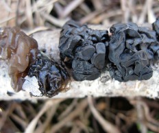
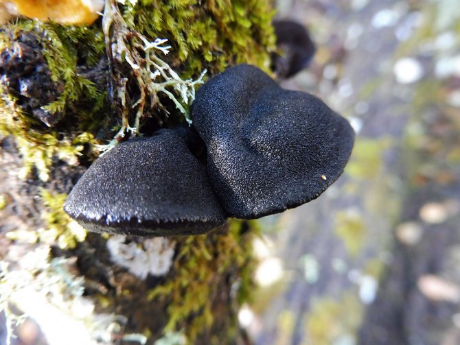
Fruiting bodies are initially rounded, tuberculate or obversely shaped, often with a tapering base, 1 - 3 cm in diameter, adherent to the substrate with a central point, developing discretely or form clusters of fruit bodies that do not merge, but retain a separate structure even in groups, dark brown or black. The surface is smooth, shiny, covered with conical tubercles. The consistency of the fruit bodies is gelatinous. When dry, the fruit bodies become hard, black and form a smoothed crust.
Microscopy
Spores 12 - 16 × 4 - 5.5 μm, allantoid, hyaline, thin-walled, with a pronounced apex and oil droplets in the protoplast.
Basidia 10 - 16 × 8 - 10 (13) μm, almost spherical or ovate, 4-spore, with a buckle at the base.
The hyphae of the tissue are branched, thin-walled, hyaline or slightly colored, with numerous buckles.
Ecology and distribution
Substance: Woody plants (living trees, bark and dead wood)
On branches and trunks of broad-leaved species, especially oak (Quercus), beech (Fagus) and hazel (Corylus).
Fruiting
The divisions correspond to the decades of the month.
Nutritional properties
Similar species
Exidia blackening (Exidia nigricans) - when groups are formed, the fruit bodies merge into a single mass.
APK "Vitus" - News

Birch dropsy (Erwinia multivora.)
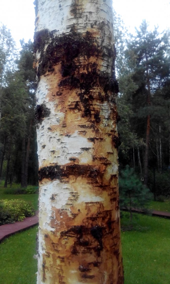
In recent years, active foci of a poorly studied disease - bacterial dropsy of birch (Erwinia multivora Scz.-Parf.), Have been identified on the territory of the Russian Federation.
Cytosporosis of birch (Cytospora horr> Posted on 21.01.2020 by the author of APK
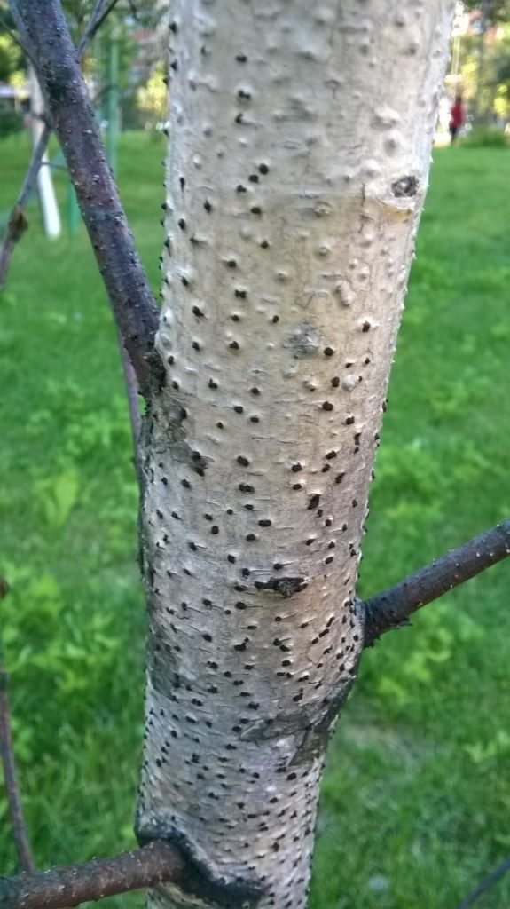
The causative agent is the mushroom Cytospora horrida Sacc. (marsupial stage - Valsa horrida Nits.), causes necrosis of the bark, drying out of individual branches and whole trees.
Exidia sugar (Ex> Published on 21.01.2020 by the author of APK
Head of the plant protection laboratory of AIC "Vitus" Sinelnikov K. Yu.
Specialists of the plant protection department of AIC "Vitus" carry out entomological and phytopathological examination of green spaces, develop individual plans for plant protection measures, treat green spaces with protective agents and carry out complex plant care.
Plant Protection Department of Agroindustrial Complex "Vitus"
8
Comparison of 3 types of exsidia: glandular exsidia, truncated exsidia and compressed exsidia
Head of the plant protection laboratory of AIC "Vitus" Sinelnikov K. Yu.
Specialists of the plant protection department of AIC "Vitus" carry out entomological and phytopathological examination of green spaces, develop individual plans for plant protection measures, treat green spaces with protective agents and carry out complex plant care.
Plant Protection Department of Agroindustrial Complex "Vitus"
8
Truncated exidia (Ex> Published on 21.01.2020 by the author of APK
Head of the plant protection laboratory of AIC "Vitus" Sinelnikov K. Yu.
Specialists of the plant protection department of AIC "Vitus" carry out entomological and phytopathological examination of green spaces, develop individual plans for plant protection measures, treat green spaces with protective agents and carry out complex plant care.
Plant Protection Department of Agroindustrial Complex "Vitus"
8
Exidia glandular (Ex> Published on 21.01.2020 by the author of APK
The causative agent is the mushroom Exidia glandulosa
Division: Basidiomycota (Basidiomycetes) Subdivision: Agaricomycotina (Agaricomycetes) Class: Agaricomycetes (Agaricomycetes) Subclass: Auriculariomycetidae Order: Auriculariales (Auriculariaceae) Family: Exidiae: Exidiaceae (Rhodes)
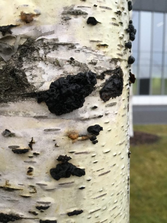
It is located on trunks with thin bark and large tree branches.
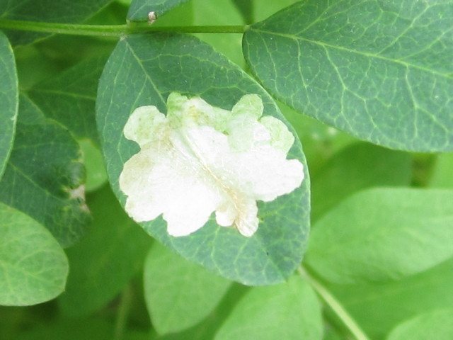
Photo: Mina acacia tail-bearing moth
Alder lurker - Cryptorrhynch> Posted on 16.12.2019 by the author of APK
a - an adult; b - an adult from the side; c - damage to the trunk with a larva; d - signs of damage to young alder; e - signs of a previous lesion on alder.
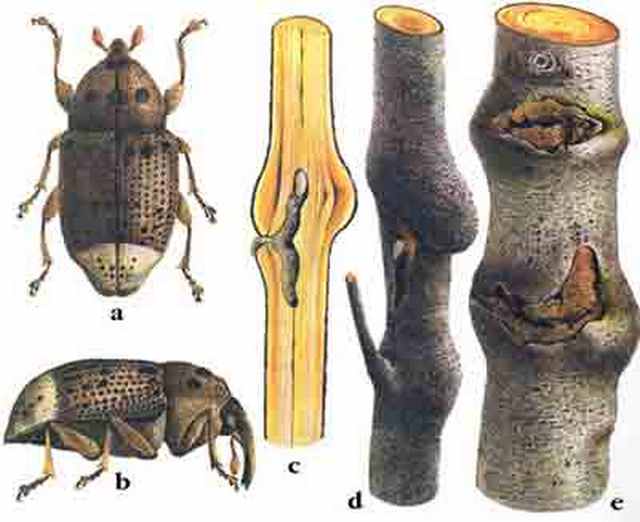
Dark-colored weevil, 6-9 mm long. The body is covered with dark brown, almost black scales with very short bristles. The shield is covered with white scales on the sides. The prothorax has a deep groove between the thighs, in which the rostrum is located. The larva is white with a brown head, legless, 10-12 mm long.
Willow diplodine necrosis (Diplodina microsperma)
Diplodine necrosis of willow trunks and branches (causative agent - the fungus Diplodina microsperma).

Photo: Sinelnikova K.Yu.
Synonyms: - Plagiostoma salicellum; - Septomyxa picea; - Cpyptodiaporthe salicella;
Diplodine necrosis of willow trunks and branches (causative agent - the fungus Diplodina microsperma) is a widespread and dangerous, but poorly understood disease. Continue reading →
Coronal sarcosphere (Sarcosphaera coronaria)
Synonyms:
- The sarcosphere is crown;
- Pink crown;
- Purple bowl;
- Sarcosphaera coronaria;
- Peziza coronaria;
- Sarcosphaera eximia.
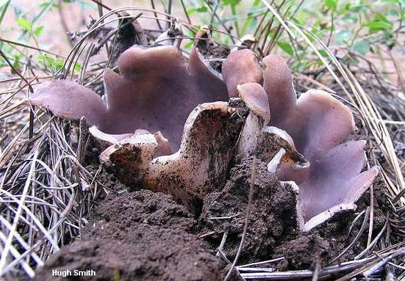
The Crown Sarcosphere (Sarcosphaera coronaria) is a mushroom of the Petsicev family, belonging to the genus of monotypic Sarcospheres.
External description
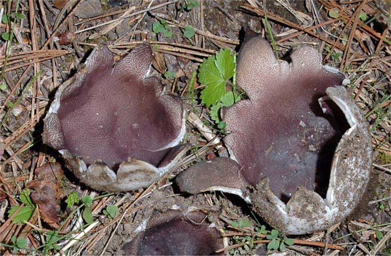
The diameter of the fruiting bodies of the coronal sarkosphere does not exceed 15 cm. Initially, they are closed, have thick walls and a spherical shape and a whitish color. A little later, they more and more protrude above the soil surface and protrude in the form of several triangular blades.
The hymen of the fungus is initially characterized by a purple color, gradually darkens more and more. On the 3-4th day after the opening of the fruit bodies, the mushroom in its appearance becomes very similar to a white flower with a very sticky surface. Because of this, the soil constantly adheres to the mushroom. The inner part of the fruiting body is wrinkled and has a purple color. From the outside, the mushroom is characterized by a smooth and white surface.
Fungal spores have an ellipsoidal shape, contain several drops of oil, are characterized by a smooth surface and dimensions of 15-20 * 8-9 microns. They have no color, in their totality they represent a white powder.
Season and habitat of the mushroom
The crown sarcosphere grows mainly on calcareous soils in the middle of forests, as well as in mountainous areas. The first fruiting bodies begin to appear in late spring, early summer (May-June). They grow well under a layer of fertile humus, and the first appearance of individual specimens falls on the time when the snow has just melted.
Edibility
There is no exact information about the edibility of the coronal sarkosphere. Some mycological experts classify this species as poisonous, others call the crown sarcosphere a pleasant and completely edible specimen of mushrooms. In the English printed sources on mycology, it is said that the corona sarcosphere mushroom should not be eaten, since there is much evidence that this type of mushroom causes severe abdominal pain, sometimes even fatal. In addition, the fruiting bodies of the crown sarcosphere are capable of accumulating toxic components, and, in particular, arsenic, from the soil.
Similar types and differences from them
The appearance of the coronal sarcosphere does not allow this species to be confused with any other mushroom. Already by the name it can be understood that the mature species has the shape of a crown, crown. This appearance makes the sarcosphere unlike other species.
Doubles and their differences
There are several types of mushrooms that are considered twins of the Compressed Exidia:
- Exidium glandular - resembles compressed in shape and color. Nevertheless, the glandular has a more saturated black color, and small warts can be seen on the surface of the fruiting body. This doppelgänger is believed to be an edible and delicious mushroom.
- Truncated exidia - similar in color and shape. You can distinguish a double from a real one by the presence of a lower velvety surface and small warts on its fruiting body. They are classified as inedible.
- Exidia blossoming - has a similar color and rounded flattened fruiting bodies. However, it will not be so difficult to distinguish a twin from a compressed exsidium, since most often it grows on a birch. This variety is never found on willow. It is an inedible species.
- Leafy shiver - similar in shape and color to fruit bodies, but this species is quite rare and grows on stumps. Experts classify it as inedible and do not recommend using it for food.
Description
Species of the genus Exidia have jelly-like mushroom bodies of irregular cerebral shape and various colors: from white transparent to black. Fruit bodies grow separately or in clusters when they can fuse. In dry weather, the mushrooms dry out, turning into hard thin crusts that remain viable for up to several years in herbarium conditions. Under natural conditions, this allows fruit bodies to withstand prolonged droughts and come to life again after rain.
In countries with a mild climate, mushrooms of this genus continue to develop continuously from early autumn to spring, so they can be attributed to winter mushrooms. Light frosts down to -10 ° C do not harm them, and during the thaw, at positive temperatures, mushrooms continue to grow and form spores. In the conditions of a more severe winter in the middle zone of the European part of Russia, fungi of some species of the genus Exidia successfully overwinter and begin to develop immediately after the snow melts. Their vital activity continues for several weeks.
Exidia
Exidia glandular Exidia compressed
Exidia is a widespread species of the large Auriculariaceae family; these mushrooms grow in Russian forests almost everywhere. They have a pulp similar to dense jelly and an indefinite shape of fruit chalk. They have a characteristic feature, they dry out in heat / drought, in cool rainy seasons they swell again and grow actively. Therefore, they become noticeable only in autumn and spring, although they can appear in cold, damp summers. In our region, two species are often found, both prefer alder-willow thickets, I have tasted (raw!) No taste at all!
Exidia glandular (Exidia glandulos)
Exidia glandular on birch
It is sometimes called glandular tremor.Actually, it does not look like mushrooms at all, fruiting bodies are born in the form of shapeless dense gelatinous tubercles densely adhered to wood. Growing up, as a rule, they merge into irregularly rounded or elongated blotches with clearly defined edges. The surface is glossy with numerous winding folds and peculiar rounded mini outgrowths (lat. Carrier glands).
Elastic flesh, odorless and absolutely tasteless. Colors of all shades of brown and black. Spores form over the entire surface of the fruiting bodies; therefore, drying mushrooms are often covered with a whitish spore powder. They grow on fallen trunks and thick branches of deciduous trees. Drying, they turn into a thin smoothed crust. Exidia glandular is edible and they write that it even has medicinal properties. Only eat raw!
Exidia compressed (Exidia recisa)
In Latin, "recisa" stands for abbreviation
Exidia compressed on aspen
Compressed exsidia mushrooms are compact, small (1 - 2.5 cm). Fruit bodies are rounded, wrinkled, with uneven edges and tiny, barely noticeable legs or a narrowed base. The outer surface is matt, the inner spore-forming is smooth and shiny. mobile pulp is translucent, thin, without any taste or smell. The color of the fruit bodies is brownish-yellow, orange-brown or reddish-brown, usually grows in small families, but usually does not form intergrowths. When dried, compressed significantly decreases in size, shrinks, becoming a dull dark brown color. Grows on decaying deciduous trees / branches, preferring willow trees. These nondescript woody mushrooms have hardly been studied, so their edibility is not known. Poisonous species are not described among exsidia, they are simply not edible due to their small size, unappetizing appearance and hard gelatinous pulp.
Sprinkled Science (Naucoria subconspersa)
Synonyms:
Alnicola subconspersa
Description
A hat with a diameter of 2-4 (up to 6) cm, convex in youth, then, with age, prostrate with a lowered edge, then flat, prostrate, possibly even slightly curved. The edges of the cap are even. The cap is slightly translucent, hygrophane, stripes from the plates can be seen. The color is light brown, yellow-brown, ocher, some sources associate the color with the color of ground cinnamon. The surface of the cap is fine-grained, fine-flaked, because of this it seems as if powdered.
The bedspread is present at a very early age, until the size of the cap does not exceed 2-3 mm, the remains of the bedspread along the edge of the cap can be found on mushrooms up to 5-6 mm in size, after which it disappears without a trace.
In the photo there are young and very young mushrooms. The diameter of the smallest cap is 3 mm. The veil is visible.
The leg is 2-4 (up to 6) cm high, 2-3 mm in diameter, cylindrical, yellow-brown, brown, watery, usually covered with a finely scaly bloom. Litter (or soil) grows from below to the leg, sprouted with mycelium, resembling white cotton wool.
The plates are not frequent, adherent. The color of the plates is similar to the color of the pulp and cap, but with age, the plates turn brown more. There are shortened plates that do not reach the stem, usually more than half of all plates.
The pulp is yellow-brown, brown, thin, watery.
The smell and taste are not pronounced.
The spore powder is brown. The spores are elongated (elliptical), 9-13 x 4-6 μm.
Habitat
It inhabits from early summer to late autumn in deciduous (mostly) and mixed forests. Prefers alder, aspen. It was also noted in the presence of willow and birch. Grows on litter or soil.
Similar species
Tubaria furfuracea is a rather similar mushroom. But it is almost impossible to confuse, since tubaria grows on wood debris, and science grows on the ground or litter. Also, in tubaria, the veil is usually more pronounced, although it may be absent. Science can only find it in very small mushrooms. Tubaria appears much earlier than science.
Science of other kinds - all science are very similar to each other, and often they cannot be distinguished without a microscope. However, the sprinkled one is distinguished by the surface of the cap, covered with fine granularity, small-scaled. Galerina sphagnorum, as well as other gallerina, for example, Galerina marsh (G. Paludosa) - in general, it is also quite similar mushroom, like all small brown mushrooms with adherent plates, however, gallerins are distinguished by the shape of the cap - similar gallerins have a dark tubercle, which is usually absent in science. Although, darkening towards the center of the cap in science is also quite common, but a tubercle is not a frequent occurrence, when it is obligatory for gallerinas, then science can rarely be, rather as an exception to the rule, and if there is, then not everyone, even in one family. Yes, and gallerinas have a smooth hat, while these sciences have fine-grained / small-scaled ones.
Edibility
Edible is unknown. And hardly anyone will check it, given the similarity with a large number of obviously inedible mushrooms, a nondescript appearance and a small amount of small fruit chalk.
Photo: Sergey
Bulgaria Inquinance
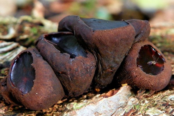
Most of the mushrooms are used for cooking various dishes, pickles, pickled delicacies, and some are only of medical interest. For example, mushrooms Bulgaria Inquinance, more like pots with strange contents, and sometimes - like a scattering of coals.
Bulgaria inquinans - lat. (Bulgaria inquinans)
In another way, this mushroom is called staining Bulgaria.
Description of the mushroom
Fruiting body
The diameter of the fruiting body reaches 10-40 mm, the height is 20 mm. Young fungi have a rounded, closed body, resembling a plaque, and held on a tiny stalk no more than 3 mm long. The rough surface of the young body is covered with tubercles and is colored ocher-brown, brown or brown-gray. The tubercles are brown - purple or brown in color.
Subsequently, in the center of the mushroom, reaching the edges, a pit with a blue, almost black bottom is formed. The mushroom itself becomes like a glass or a depressed pot, which turns into a saucer with old age.
At the top of the staining Bulgaria there is a glossy disc that turns brown - red, bluish - black, and then - in olive - black tone. On fine days, it remains smooth and dull, in rainy weather it damp and shines. Old mushrooms shrivel and dry out.
The mushroom is filled with a jelly-like, tight, ocher-brown flesh that exudes a pleasant mushroom aroma or does not smell at all.
Bulgaria Inquinance reproduces by black and brown smooth kidney-shaped spores contained in black spore powder... They are abundantly isolated from mushrooms and stain the nearby soil in a black-brown hue, for which they were called dirty.
Growing places
For growth, the fungus chooses dead wood and other remains of oak, aspen, birch, beech and other trees located in mixed and deciduous forests, in park areas. Prefers shaded areas and appears in large numbers after rains. Often found in North American and European countries.
Group fruiting in Bulgaria, which stains, depending on climatic characteristics, falls on July - November.
Edibility
Although a pleasant aroma often emanates from this mushroom, Bulgaria is not eaten because of its specific type and tasteless pulp.
Similar types and differences from them
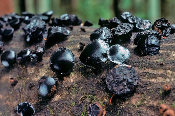
Bulgaria Inquinance can be confused with the following inedible species:
- The exidia is truncated. Prefers dead oak branches and grows in crooked black fruiting chalk.
- The sarcosoma is spherical. It differs in less group fruiting (no more than three fruiting bodies are combined together) and chooses coniferous forests for growth.
Healing properties
This Bulgaria contains many sugars, quinones, proteins and specific lactones, as well as fatty acids (oleic, palmitic, oxalic, phthalic, etc.) and eighteen amino acids. Thanks to such a rich composition, this mushroom has a lot of medicinal properties:
- Suppresses the growth of cancerous tumors. For example, sarcoma-180 cells are destroyed by 60 percent of the mushroom extract.At the same time, immunity decreases slightly, in contrast to the use of chemotherapy, which kills a lot of healthy cells.
- Improves the rheological parameters of blood cells. Due to some diseases, blood does not move well through the vessels, blood circulation is disturbed, the blood thickens, the shape of erythrocytes is disturbed. The alcoholic extract of the mushroom improves the condition of blood cells and the speed of their movement through the vessels.
Despite the presence of these medicinal properties, Bulgaria Inquinance is not recommended for treatment during pregnancy and lactation.
What does exidium glandular look like?
The description of the glandular exsidia must begin with the fruiting body. It is low, reaching a height of 1-2 cm. Outside, it is black. Inside is a transparent or olive brown jelly-like substance. The young mushroom has a teardrop shape. Having grown, it acquires a fruiting body, similar to the structure of the human brain: tuberous and ear-shaped.
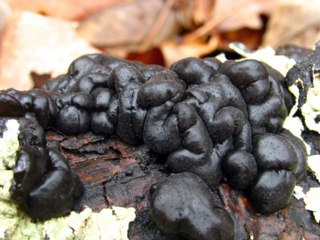
When dry, the color becomes dull. The body hardens to form a dense crust. With increasing humidity, it returns to its original state. By consistency - soft density, similar to swollen gelatin or marmalade. Adult plants form a continuous colony, growing together into a single whole. Odorless. The taste is weak. Other structural features:
- The fruits of the mushroom are white, curved, cylindrical in shape. Disputes are produced all year round (in winter - during warming).
- The hypha (mushroom web) is branched and equipped with buckles.
- The reproductive organs (basidia) are in the form of a ball or egg and form 4 spores each.

